Development and Optimization of a pH-Sensitive Fluorescence-Based Assay for Quantitative Antibody Internalization Analysis
 Publication Date:2026-07-31
 Page Views:14
Overview
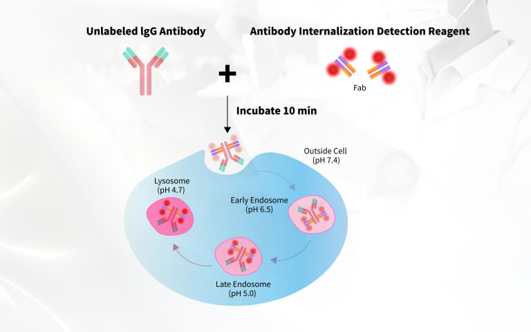
Antibody internalization analysis is widely used to characterize antibody behavior after target binding and provides important information for antibody discovery and antibody-drug conjugate (ADC) development. A key step in the mechanism of action (MOA) of ADCs is receptor-mediated endocytosis. Upon binding to its target antigen on the cell surface, the ADC-antigen complex is internalized through endocytic pathways, forming early endosomes. The early endosomes are subsequently trafficked to late endosomes and lysosomes, where the linker is cleaved—either enzymatically or chemically—to release the active cytotoxic drug. The released payload then exerts its anti-tumor activity, typically by disrupting DNA replication or microtubule function, leading to cell apoptosis or death.1,2
Given this mechanism, efficient internalization of the ADC is a prerequisite for therapeutic efficacy. The internalization process is influenced by several factors, including antigen density on the cell surface, antibody affinity for the antigen, and epitope accessibility. Therefore, screening antibodies with high-efficiency internalizing capabilities is critical during early-stage development.3
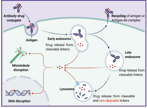
Figure 1. Classic mechanism of action of ADC drugs.2
Endocytosis assessment is necessary across multiple stages of ADC development:
- ✔ Discovery stage: To identify antibodies that not only bind specifically to tumor-associated antigens but also internalize efficiently.
- ✔ Lead optimization: To evaluate how variations in antibody structure or conjugation chemistry affect internalization and intracellular trafficking.
- ✔ Preclinical development: To confirm that the selected ADC candidate exhibits consistent internalization behavior across relevant cell models.
- ✔ Mechanism-of-action studies: To validate the intracellular delivery of the payload and its correlation with cytotoxic effects.
The main purposes of measuring endocytosis include quantitative analysis of ADC internalization rate, analysis for intracellular localization of ADC, and high-throughput screening of candidate ADC molecules. Over the years, several techniques have been used to study antibody internalization including radiolabels, fluorescent microscopy, flow cytometry and cellular toxicity assays.4
Multiple analytical approaches are available for investigating ADC internalization, each offering distinct advantages: live-cell imaging facilitates real-time visualization of fluorescently labeled ADCs, radioactive labeling enables accurate quantification of antibody uptake, electron microscopy provides nanometer-scale localization details, while toxin-based assays establish a direct correlation between internalization and cytotoxic activity, delivering functionally relevant data.
In this article, we present a newly developed endocytosis detection reagent designed for sensitive and reliable measurement of antibody internalization in both HER2+ adherent cells and CD20-positive suspension cells. We explore key experimental parameters, including dye concentration and incubation time, and evaluate their impact on fluorescence signal and internalization efficiency.
Methods and Materials
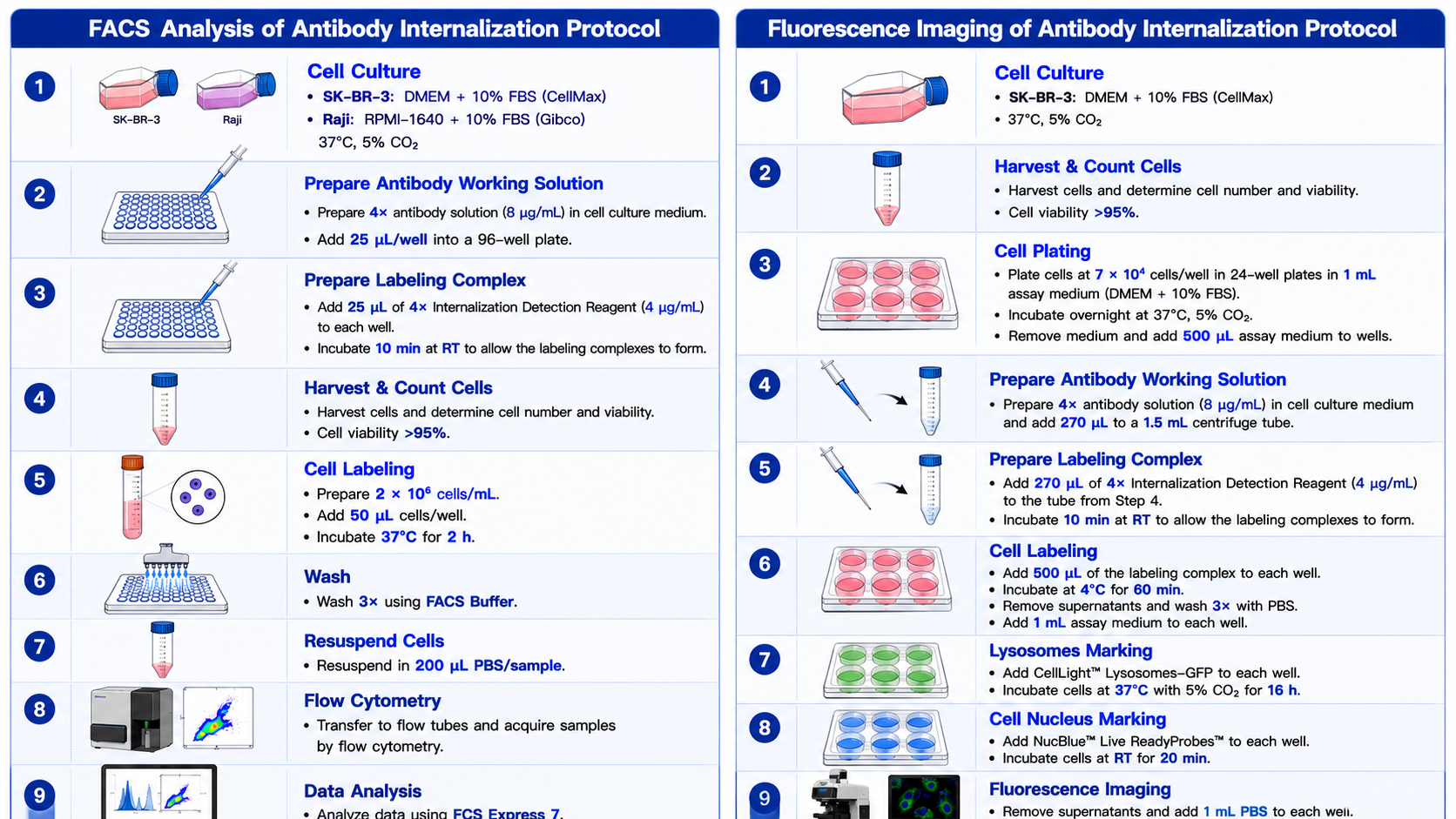
Lyophilized reagents were reconstituted using sterile deionized water into the mother solution (200 μg/mL) and diluted into 4× working solution (4 μg/mL) before use.
Results and Discussion
Specificity Validation of the Internalization Detection Reagent
To validate the specificity of the internalization detection reagent, we performed detection in HER2+ cell lines and used the HER2-specific antibody Trastuzumab Biosimilar as well as a nonspecific IgG1 isotype control. Fluorescence microscopy revealed bright fluorescent signals exclusively in the endosomal compartments of test group cells (Fig. 2C), while control groups (either antibody-free or with isotype control antibody) showed minimal background signal (Fig. 2A–B). This antibody-dependent fluorescence pattern clearly demonstrates that the internalization signal is specifically mediated by target antibody binding. Z-stack scanning further confirmed this conclusion, demonstrating high colocalization between the internalization reagent signal (Red) and lysosomal markers (Green), with no extracellular signal detected (Fig. 2D).
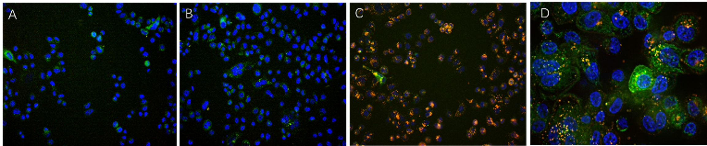
Figure 2. SK-BR-3 cells were treated with CellLights Lysosome GFP (green) for 16 hours followed by treatment with Anti-Her2 Abs-Internalization Detection Reagent conjugate and IgG1 Isotype-Internalization Detection Reagent conjugate separately for 16 hours (red), then stained with NucBlue Live ReadyProbes (blue) for 20 minutes and imaged on the EVOS M7000. A. Antibody Internalization Detection Reagent (IGG-PZF2001). B. IgG1 Isotype-Internalization Detection Reagent conjugate. C. Anti-Her2 Abs-Internalization Detection Reagent conjugate. D. Anti-Her2 Abs-Internalization Detection Reagent conjugate (Z-stacking).
Furthermore, flow cytometric analysis was performed in both HER2+ and CD20+ cell lines. The results demonstrated that this internalization detection reagent is not only applicable for antibody internalization assessment across multiple cell types, but also enables rapid and quantitative analysis through flow cytometry (Fig. 3A–B).
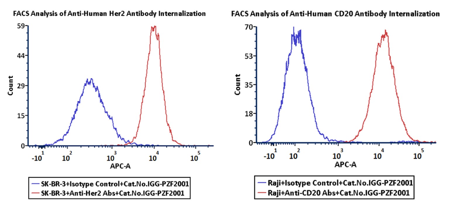
Figure 3. FACS analysis of Antibody Internalization. A. HER2 specific antibody Trastuzumab (Red) in HER2+ cell line. B. CD20 specific antibody Rituximab (Red) in CD20+ cell line. Blue: IgG1 isotype control.
Optimization of Internalization Detection Reagent Concentration
To establish the optimal working concentration, we systematically evaluated the internalization detection reagent in CD20+ and HER2+ cell lines using a fixed primary antibody concentration of 2 μg/mL while detection reagent concentrations of 1 μg/mL, 2 μg/mL, and 4 μg/mL. Dose-dependent analysis revealed that increasing the detection reagent concentration from 1 μg/mL to 4 μg/mL significantly enhanced positive signals, while higher concentrations (4 μg/mL) concomitantly increased background signals in control groups. Through comprehensive signal-to-noise ratio (SNR) analysis, we determined that a 1:2 ratio (1 μg/mL detection reagent to 2 μg/mL antibody) provided the optimal balance between detection sensitivity and background suppression in both cell lines (Fig. 4A–D). This optimized condition establishes a reliable and cost-effective protocol for subsequent internalization assays.
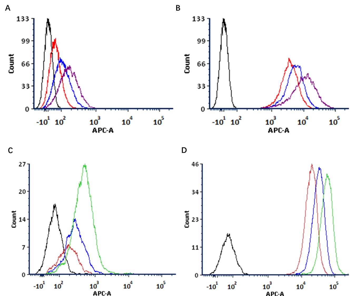
Figure 4. FACS analysis of Antibody Internalization with different detection reagent concentrations. A: Detection Reagent control in CD20+ cell line. B: Detection Reagent and CD20 specific antibody Rituximab in CD20+ cell line. Blank: Only cell. Red: 1 μg/mL Detection Reagent, Blue: 2 μg/mL Detection Reagent, Purple: 4 μg/mL Detection Reagent. C: Detection Reagent control in HER2+ cell line. D: Detection Reagent and HER2 specific antibody Trastuzumab in HER2+ cell line. Blank: Only cell. Red: 1 μg/mL Detection Reagent, Blue: 2 μg/mL Detection Reagent, Green: 4 μg/mL Detection Reagent.
Temporal Dynamics of Antibody Internalization
We investigated the kinetic profiles of antibody internalization by incubating the antibody-reagent complex with CD20+ and HER2+ cell lines for 30 minutes to 4 hours followed by flow cytometric analysis. The results demonstrated cell type-dependent internalization kinetics, with CD20+ cells showing a linear increase in fluorescence intensity throughout the time course, while HER2+ cells exhibited a biphasic response characterized by minimal signal change during the initial 2 hours followed by a significant increase at 4 hours (Fig. 5A–D).
These differential kinetic patterns confirm that our internalization detection reagent can reliably monitor the dynamic internalization process through quantitative fluorescence changes while revealing target-specific variations in internalization rates. The findings underscore the importance of establishing cell line-specific detection windows, as optimal timepoints may vary significantly depending on both the target antigen and cellular context. This temporal characterization provides critical guidance for experimental design in future applications, emphasizing that internalization kinetics should be empirically determined for each new antibody-cell system combination to ensure accurate assessment of internalization efficiency.
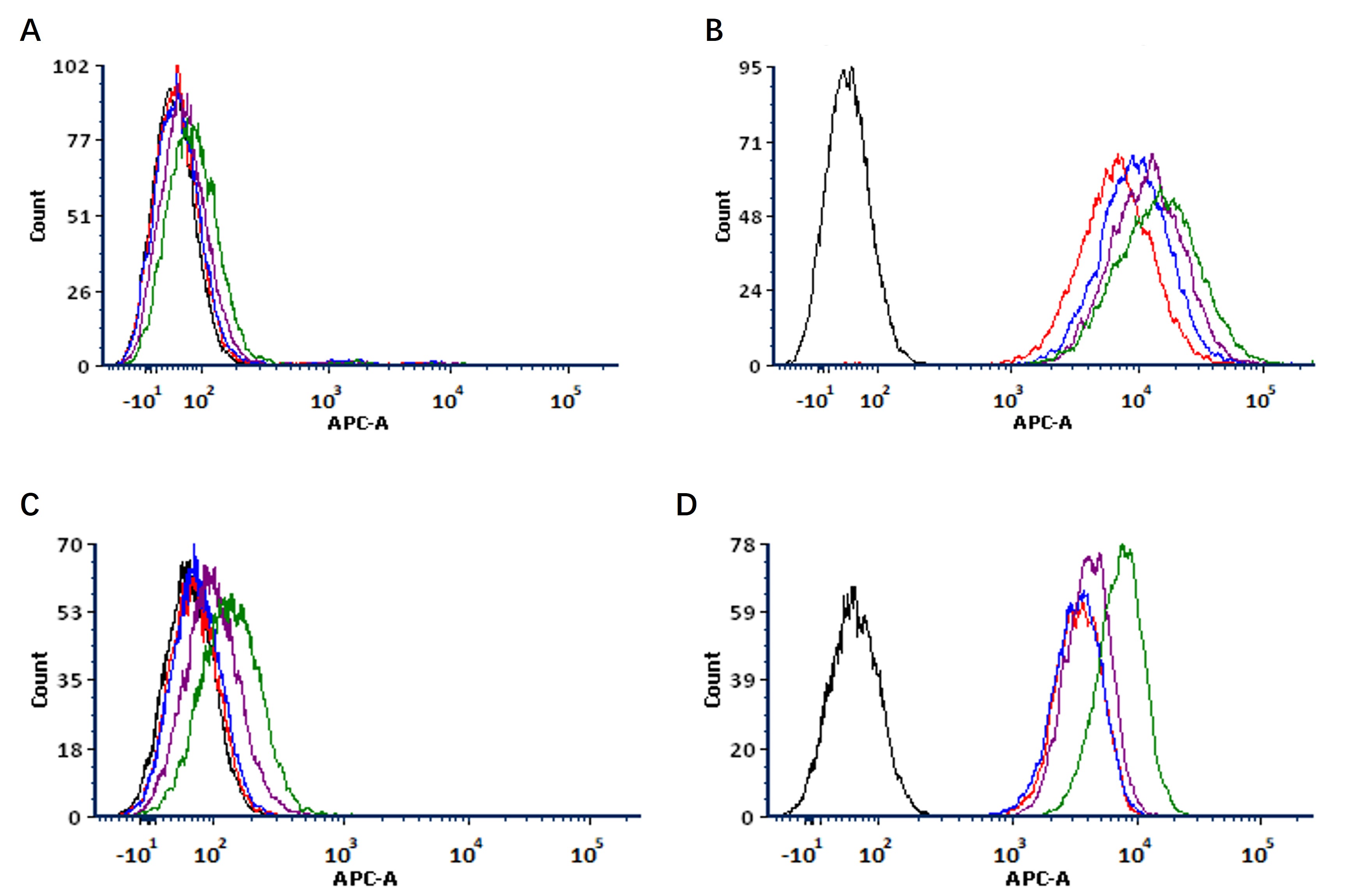
Figure 5. FACS analysis of Antibody Internalization with different incubation times. A: Detection Reagent control in CD20+ cell line. B: Detection Reagent and CD20 specific antibody Rituximab in CD20+ cell line. C: Detection Reagent control in HER2+ cell line. D: Detection Reagent and HER2 specific antibody Trastuzumab in HER2+ cell line. Blank: Only cell. Red: 30 min, Blue: 1 h, Purple: 2 h, Green: 4 h.
Conclusion
The field of antibody-drug conjugates (ADCs) has seen remarkable advancements in recent years, with multiple ADC therapies gaining regulatory approval. The therapeutic efficacy of ADCs in oncology stems from their ability to selectively internalize into target cells and release potent payloads within the tumor microenvironment. A critical factor in ADC development is the establishment of robust, high-throughput methods to assess antibody internalization efficiency. We have developed a pH-sensitive fluorescent dye-based internalization assay, which demonstrates strong signal amplification in acidic endosomal compartments while maintaining minimal background in both HER2+ and CD20+ cell models.
ACROBiosystems pHintra™ Internalization Detection Solution
pHintra™ Internalization Detection Solution supports antibody internalization analysis by both flow cytometry and fluorescence imaging across all stages of ADC research—from antibody discovery to lead optimization and preclinical characterization. The reagent has been validated in both adherent and suspension cell models and is compatible with diverse antibody programs, providing a standardized and scalable workflow for quantitative internalization assessment.
Explore pHintra™ Internalization Detection Solution
Frequently Asked Questions (FAQ)
Q1: What is antibody internalization, and why is it important for ADC development?
A: Antibody internalization refers to the process by which an antibody–antigen complex is internalized into cells after binding to a target on the cell surface. For antibody-drug conjugates (ADCs), efficient internalization is essential because it enables intracellular trafficking to endosomes and lysosomes, where payload release occurs. As a result, internalization assessment has become an important component of antibody screening, ADC lead optimization, and mechanism-of-action studies, helping researchers identify candidates with efficient intracellular delivery.
Q2: At what stage of ADC development should antibody internalization be evaluated?
A: Antibody internalization can provide valuable insights throughout ADC development. During antibody discovery, it helps prioritize candidates with efficient cellular uptake. During lead optimization, it supports comparison of antibody variants, linker designs, or conjugation strategies that may affect intracellular trafficking. In preclinical studies, it also helps characterize mechanisms of action and confirm consistent uptake across target-expressing cell models. pHintra™ Internalization Detection Solution supports antibody internalization analysis by both flow cytometry and fluorescence imaging across these research stages.
Q3: Is strong antigen binding always associated with efficient antibody internalization?
A: No. High binding affinity does not necessarily translate into efficient internalization. Cellular uptake is influenced by multiple factors, including epitope accessibility, receptor recycling, antigen density, and endocytic pathways. Therefore, antibodies with similar binding characteristics may exhibit markedly different internalization behaviors. Combining binding assays with internalization analysis provides a more comprehensive evaluation during antibody selection. pHintra™ Internalization Detection Solution enables direct assessment of antibody internalization to complement binding studies.
Q4: How should researchers choose between flow cytometry and fluorescence imaging for antibody internalization studies?
A: The choice depends on the experimental objective. Flow cytometry is ideal for rapid, quantitative comparison of antibody internalization across large cell populations, whereas fluorescence imaging reveals intracellular localization and trafficking. These approaches are complementary and are often used together during antibody screening and ADC research.
Q5: Can one antibody internalization assay workflow be applied across different antibody targets and cell models?
A: Yes, although assay parameters such as antibody concentration, incubation time, and cell density should be optimized for individual targets and cell types. A standardized detection workflow can nevertheless be applied across diverse antibody programs, improving experimental consistency and simplifying assay development. pHintra™ Internalization Detection Solution has been validated in both adherent and suspension cell models and is compatible with flow cytometry and fluorescence imaging.
References
1. Shivatare VS, Huang HW, Tseng TH, Chuang PK, Zeng YF, Wong CH. Probing the Internalization and Efficacy of Antibody-Drug Conjugate via Site-Specific Fc-Glycan Labelling of a Homogeneous Antibody Targeting SSEA-4 Bearing Tumors. Isr J Chem. 2023 Oct;63(10-11):e202300042.
2. Fu Z, Li S, Han S, et al. Antibody drug conjugate: the "biological missile" for targeted cancer therapy. Sig Transduct Target Ther 7, 93 (2022).
3. ProBioCDMO. ADC bioassay services. https://www.probiocdmo.com/add-adc-bioassay-service.html
4. Nath N, Godat B, Zimprich C, Dwight SJ, Corona C, McDougall M, Urh M. Homogeneous plate based antibody internalization assay using pH sensor fluorescent dye. J Immunol Methods. 2016 Apr;431:11-21.
 Popular ArticlesRelated RecommendationsPopular Events
 Reliable Antibody Internalization Assessment: Approaches and Considerations for Antibody Discovery and ADC Research2026-07-31Page Views:19
.jpg) Development and validation of bioanalytical assays for the quantification of ADC2026-06-17Page Views:354
Development and validation of bioanalytical assays for the quantification of ADC2026-06-17Page Views:354 Dual-Payload ADCs: Translational Challenges and Clinical Pharmacology2026-06-16Page Views:423
 Next-Generation Dual-Payload ADCs: Target Selection, Payload Synergy, and Clinical Pipeline Analysis2026-06-12Page Views:3006
382-240.jpg) B7-H3 (CD276): Clinical Progress and Emerging Therapeutic Strategies in Solid Tumors2026-05-26Page Views:1158
B7-H3 (CD276): Clinical Progress and Emerging Therapeutic Strategies in Solid Tumors2026-05-26Page Views:1158




 Related Products