Save Product

Product Details
Synonyms
IGF-I R / IGF-1 R
Source
Biotinylated Rhesus macaque IGF-I R Protein, His Tag, primary amine labeling (IGR-R82H4) is expressed from human 293 cells (HEK293). It contains AA Glu 31 - Asn 932 (Accession # I2CWY3). It is the biotinylated form of Rhesus macaque IGF-I R Protein, His Tag (IGR-R52H3). It is the biotinylated form of Rhesus macaque IGF-I R Protein, His Tag (Cat. No. IGR-R52H3).
Predicted N-terminus: Glu 31 & Asp 741
Request for sequenceMolecular Characterization

Other Tags and Version Biotin & Other Labeled Version
This protein carries a polyhistidine tag at the C-terminus.
The protein has a calculated MW of 81.1 kDa & 23.8 kDa. The protein migrates as 95-130 kDa when calibrated against Star Ribbon Pre-stained Protein Marker under reducing (R) condition (SDS-PAGE) due to glycosylation.
The protein is designed as a dimer.
Labeling
The primary amines in the side chains of lysine residues and the N-terminus of the protein are conjugated with biotins using standard chemical labeling method. A standard biotin reagent (13.5 angstroms) is used in this product.
Protein Ratio
Passed as determined by the HABA assay / binding ELISA.
Purity
>90% as determined by SDS-PAGE.
Formulation
Lyophilized from 0.22 μm filtered solution in PBS, pH7.4 with trehalose as protectant.
Contact us for customized product form or formulation.
Reconstitution
Please see Certificate of Analysis for specific instructions.
For best performance, we strongly recommend you to follow the reconstitution protocol provided in the CoA.
Storage
For long term storage, the product should be stored at lyophilized state at -20°C or lower.
Please avoid repeated freeze-thaw cycles.
This product is stable after storage at:
- -20°C to -70°C for 12 months in lyophilized state;
- -70°C for 3 months under sterile conditions after reconstitution.
ACRO Quality Management System
Customers Also Viewed
Performance Data
SDS-PAGE
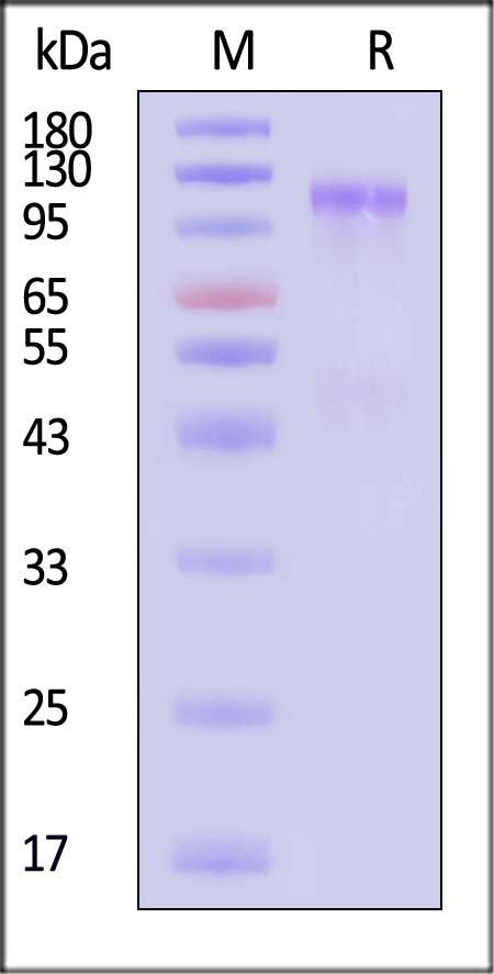
Biotinylated Rhesus macaque IGF-I R Protein, His Tag, primary amine labeling on SDS-PAGE under reducing (R) condition. The gel was stained with Coomassie Blue. The purity of the protein is greater than 90% (With Star Ribbon Pre-stained Protein Marker).
SEC-MALS
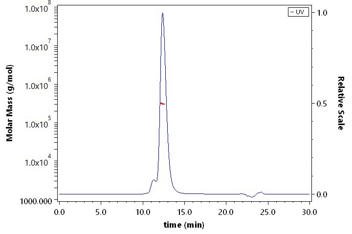
The purity of Biotinylated Rhesus macaque IGF-I R Protein, His Tag, primary amine labeling (Cat. No. IGR-R82H4) is more than 85% and the molecular weight of this protein is around 285-325 kDa verified by SEC-MALS.
Report
Bioactivity-ELISA
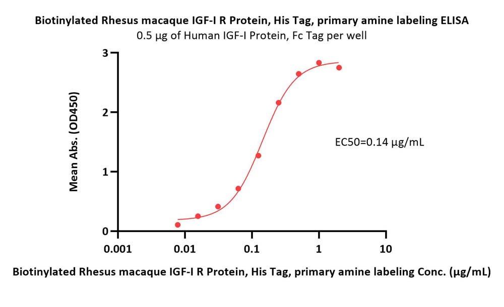
Immobilized Human IGF-I Protein, Fc Tag (Cat. No. IG1-H5263) at 5 μg/mL (100 μL/well) can bind Biotinylated Rhesus macaque IGF-I R Protein, His Tag, primary amine labeling (Cat. No. IGR-R82H4) with a linear range of 0.008-0.25 μg/mL (QC tested).
Protocol
Customer Reviews Writing Reviews

 Contact Us


 Drug Candidate Licensing