



 Nov 17, 2026
 Page Views:92


Glutamate is the principal excitatory neurotransmitter in the central nervous system (CNS), mediating fast synaptic transmission. However, when extracellular glutamate homeostasis is disrupted, this essential signaling molecule can trigger excitotoxicity, a pathological process implicated in stroke, Alzheimer’s disease (AD), multiple sclerosis, and other neurological disorders. Understanding this transition is essential for developing neuroprotective therapies, yet accurately modeling human excitotoxicity remains a major challenge in translational research.
The Dual Role of Glutamate: From Physiological Signaling to Excitotoxicity
Under normal conditions, glutamate signaling is mediated by two receptor families: ionotropic glutamate receptors, including AMPA, kainate, and NMDA receptors, which mediate fast synaptic transmission, and metabotropic glutamate receptors (mGluR1–8), which regulate neuronal activity through G-protein-coupled pathways 1,2. Together, these pathways support synaptic plasticity, learning, and memory3.
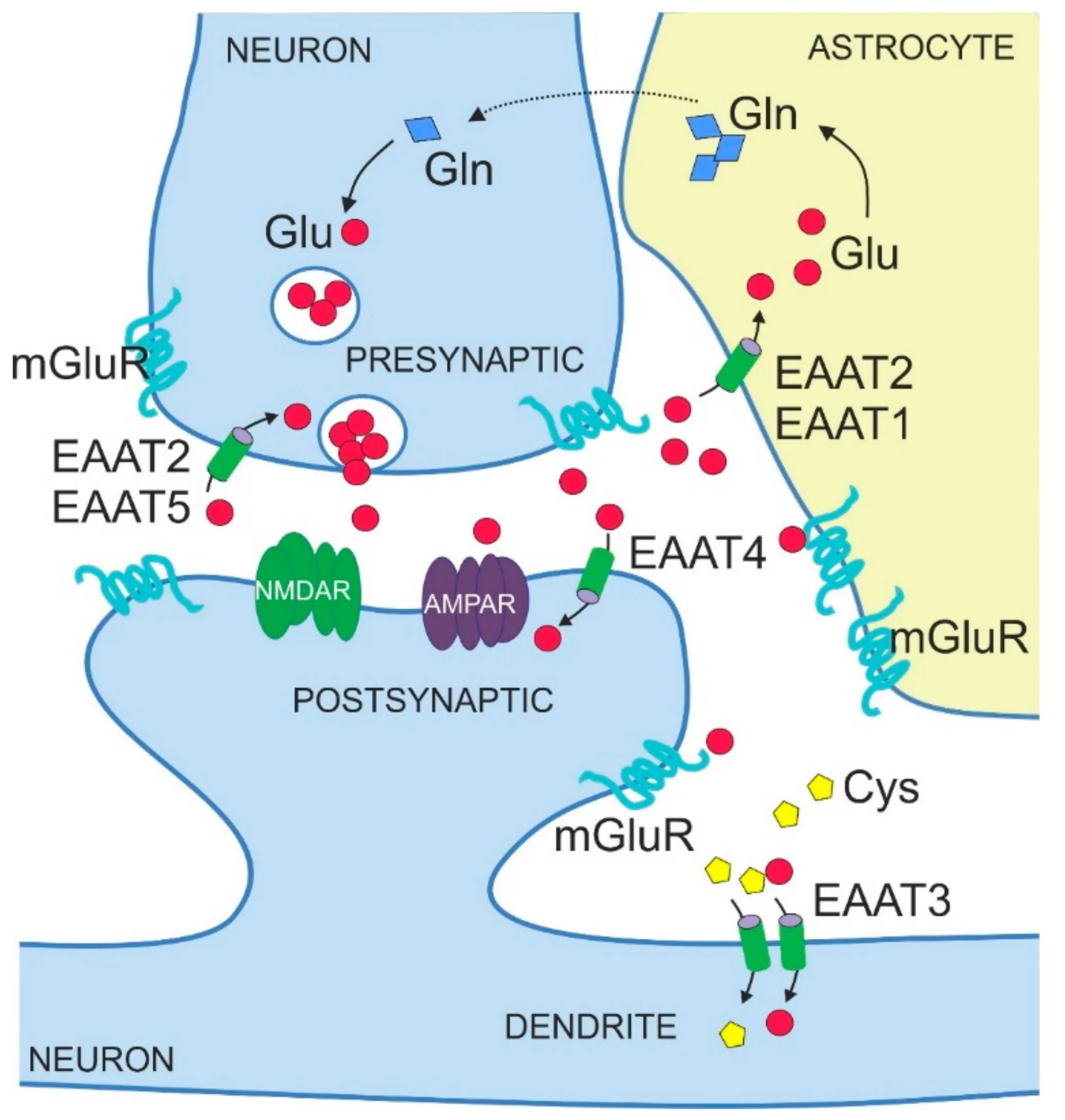
https://doi.org/10.3390/ijms20225671
Figure 1. Schematic illustration of glutamate homeostasis
For glutamate to act as a neurotransmitter, extracellular glutamate levels must be kept under strict control. After its release from presynaptic terminals, extracellular glutamate must be rapidly cleared to prevent excessive receptor activation. This process is primarily mediated by excitatory amino acid transporters (EAATs), particularly EAAT1/GLAST and EAAT2/GLT-1, which are predominantly expressed by astrocytes. These transporters also support the glutamate–glutamine cycle between neurons and astrocytes. When glutamate uptake is impaired, extracellular glutamate can accumulate and contribute to excitotoxicity ⁴.
How Excitotoxicity Develops: From Calcium Overload to Neuronal Death
NMDA receptors (NMDARs) play a central role in both synaptic plasticity and excitotoxicity due to their voltage-dependent Mg²⁺ block and high Ca²⁺ permeability 1,2. Excessive receptor activation can cause intracellular Ca²⁺ overload, initiating a cascade of cellular damage:
- Excessive receptor activation: Elevated extracellular glutamate overactivates postsynaptic NMDARs and AMPARs, driving excessive Ca²⁺ and Na⁺ influx and disrupting ionic homeostasis ⁴.
- Mitochondrial dysfunction and oxidative stress: Intracellular Ca²⁺ overload impairs mitochondrial function, reduces ATP production, and promotes the generation of reactive oxygen species (ROS) 4,5.
- Activation of cell death pathways: Elevated Ca²⁺ activates enzymes such as calpain, phospholipase A2 (PLA2), and neuronal nitric oxide synthase (nNOS), contributing to cytoskeletal damage, lipid peroxidation, and the initiation of cell death programs 4,5.
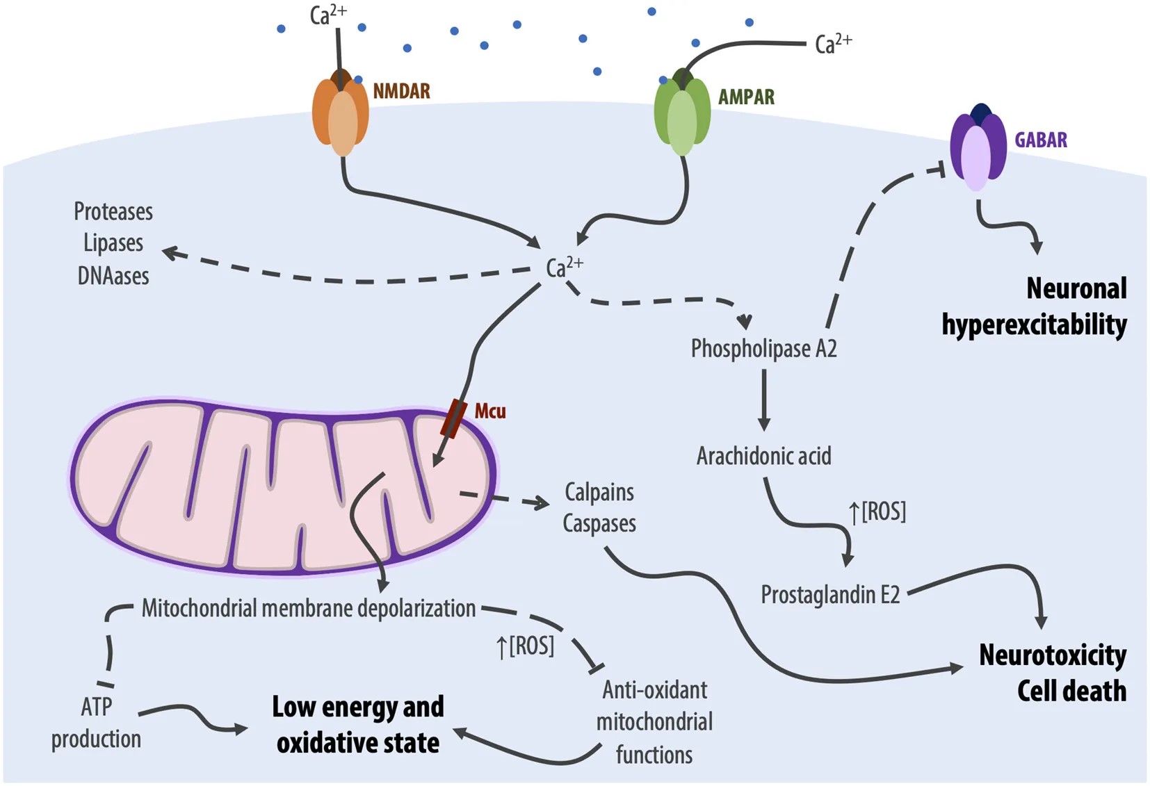
https://doi.org/10.3389/fncel.2020.00090
Figure 2. Schematic illustration of excitotoxicity
Research has also highlighted the distinct signaling roles of synaptic and extrasynaptic NMDARs. Synaptic NMDAR activation, often associated with GluN2A-containing receptors, can support neuroprotective signaling involving CREB and BDNF. In contrast, excessive extrasynaptic NMDAR activation, frequently associated with GluN2B-containing receptors, can engage pro-death pathways ⁶. This distinction helps explain why broad NMDAR blockade has faced challenges in clinical development: it can interfere with both physiological and pathological signaling.
Memantine, an uncompetitive NMDAR antagonist approved for moderate-to-severe AD, can preferentially inhibit excessive NMDAR activity and has informed research into extrasynaptic NMDAR signaling ⁷. However, the distinction is not absolute, as both synaptic and extrasynaptic NMDARs contribute to normal neuronal function. The effects of receptor activation depend on factors including receptor composition, activation intensity, and duration 8,9.
When Glutamate Becomes Harmful: Acute Injury and Chronic Neurodegeneration
Ischemic stroke is a classic example of acute excitotoxicity. During ischemia, oxygen and glucose deprivation rapidly depletes ATP, impairing ion transport and causing membrane depolarization. Glutamate is released in excess, while energy failure can also reverse the direction of glutamate transport, further increasing extracellular glutamate levels. This triggers excessive receptor activation and neuronal injury ⁵. Despite strong preclinical evidence, NMDAR antagonists have repeatedly failed to demonstrate clinical efficacy in stroke trials, highlighting the challenges of translating findings from animal models to patients ⁷.
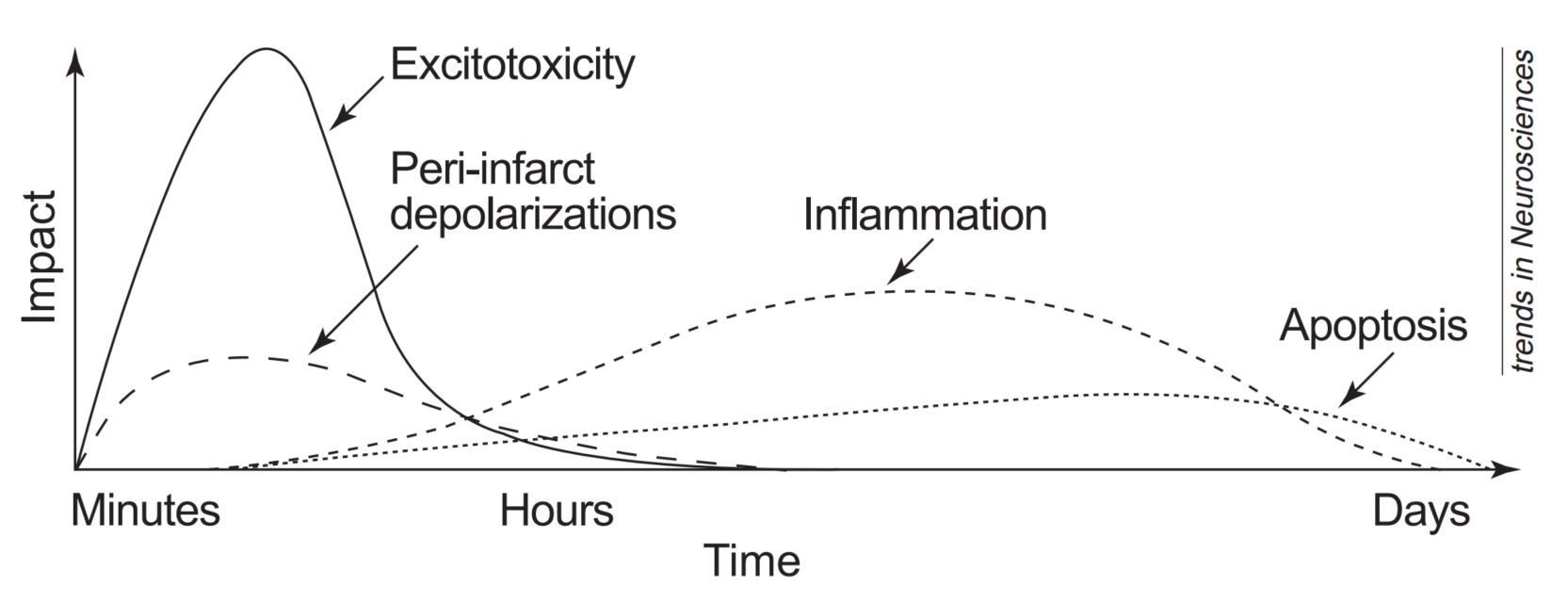
https://doi.org/10.1016/S0166-2236(99)01401-0
Figure 3. Excitotoxicity damages neurons early in cerebral ischemia and drives the downstream cascade
In Alzheimer’s disease, excitotoxicity develops more gradually. Amyloid-β (Aβ) oligomers can disrupt glutamate homeostasis by impairing astrocytic EAAT2 function and enhancing extrasynaptic NMDAR signaling. The resulting sustained increase in glutamatergic activity can impair synaptic function and exacerbate mitochondrial dysfunction and neuroinflammation, contributing to synapse loss and neurodegeneration 7,10.
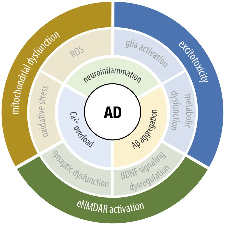
https://doi.org/10.3389/fncel.2020.00090
Figure 4. Interplay between upstream pathology and phenotype in AD
Excitotoxicity has also been implicated in multiple sclerosis, where it contributes to oligodendrocyte injury and demyelination; epilepsy, where excessive glutamatergic activity is associated with neuronal hyperexcitability and injury; and glaucoma, where it may contribute to retinal ganglion cell degeneration ¹¹⁻¹³.
Brain Organoids: A Human-Relevant Model for Excitotoxicity Research
Drug development targeting glutamate-induced excitotoxicity has long been limited by the lack of physiologically relevant models. Conventional 2D primary cultures lack three-dimensional architecture and multicellular interactions, while rodent models differ from human systems in aspects such as NMDAR subunit composition and developmental timing.
ACROBiosystems’ ready-to-use hiPSC-derived dorsal forebrain organoids provide a human-relevant 3D model for investigating glutamatergic signaling and neuronal overexcitation. Key validated features include:
- Glutamatergic neuronal composition: By days 57–87 of differentiation, the organoids contain vGLUT1⁺ glutamatergic neurons, alongside CTIP2⁺ deep-layer cortical neurons and MAP2⁺ mature neurons.
- Multicellular composition: GFAP⁺ astrocytes and OLIG2⁺ oligodendrocyte-lineage cells provide opportunities to investigate multicellular interactions involved in glutamate homeostasis, including astrocyte-mediated glutamate uptake.
- Functional validation: Microelectrode array (MEA) recordings demonstrate spontaneous network activity in all five organoids tested (5/5), while FLIPR calcium imaging reveals spontaneous rhythmic Ca²⁺ transients. Both glutamate and Yoda1 induce neuronal overexcitation, accompanied by increased firing rates and active electrode counts. These responses support the use of the organoids to study neuronal overexcitation and evaluate drug-induced changes in neuronal activity.
Product list
|
Cat. No. |
Product Description |
Product Size |
|---|---|---|
|
Ready-to-use hiPSC-Derived Dorsal Forebrain Organoids (Standard) |
5 / 20 / 100 organoids |
|
|
Ready-to-use hiPSC-Derived Dorsal Forebrain Organoids (Mature) |
5 / 20 / 100 organoids |
|
|
hiPSC-Derived Dorsal Forebrain Organoid Maintenance Kit |
60 / 120 / 360 mL |
From physiological signaling to excitotoxicity, glutamate plays a critical role in neuronal health and disease. Human iPSC-derived brain organoids offer researchers a human-relevant 3D model to investigate this transition and evaluate potential interventions, supporting the development of neuroprotective therapies.