


 Nov 9, 2026
 Page Views:76


While single-target IL-17A inhibitors remain the clinical mainstream, the dual-target IL-17A& IL-17F paradigm is rapidly advancing worldwide. Bimekizumab is now approved across psoriasis, PsA, ankylosing spondylitis, and HS. In March 2026, the BE BOLD head-to-head trial made it the first biologic to demonstrate superiority over an IL-23 inhibitor (risankizumab/Skyrizi®) in PsA, achieving ACR50 response in 49.1% vs. 38.4% at Week 16 (p=0.0078).UCB in the U.S. In BE HEARD I/II, HS patients maintained clinical responses through 3 years, with continued improvements in HiSCR response rates. Single-target and dual-target approaches are advancing in parallel, reflecting the current landscape of autoimmune drug development. Regardless of the strategy, reliable biomarker assays are essential for precisely tracking target dynamics, evaluating therapeutic efficacy, and identifying the patients most likely to benefit.
IL-17A and IL-17F: Why Dual Targeting Matters
IL-17A and IL-17F share approximately 50% amino acid sequence identity. Both cytokines are produced primarily by Th17 cells and can form three distinct dimers: IL-17A/A and IL-17F/F homodimers, and the IL-17A/F heterodimer (Figure 1). Like the homodimers, the IL-17A/F heterodimer signals through the IL-17RA/IL-17RC receptor complex. However, its interactions with the receptor subunits differ from those of the individual homodimers. Structural studies have shown that IL-17RA can engage either the IL-17A or IL-17F subunit of the heterodimer with comparable affinity, providing a structural rationale for differentiated approaches to ligand blockade [1]. IL-17A generally exhibits greater proinflammatory potency than IL-17F and has consequently been the primary focus of many approved therapies targeting this pathway. However, the relatively lower intrinsic activity of IL-17F does not necessarily translate into a lesser role in disease pathogenesis. Emerging evidence suggests that its contribution to chronic inflammation has been substantially underestimated, underscoring the therapeutic rationale for targeting both IL-17A and IL-17F.
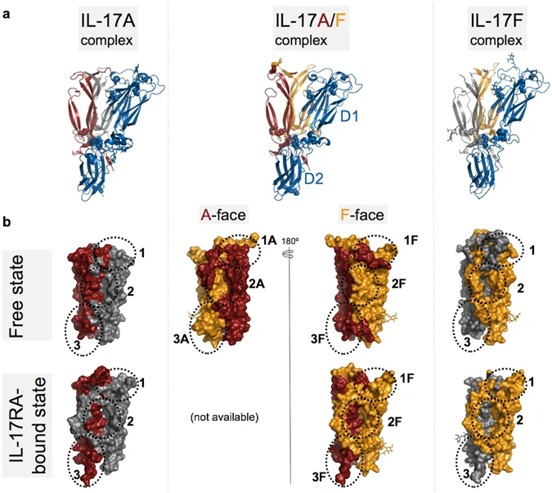
Figure 1. Structural comparison of IL-17RA-cytokine complexes.
https://doi.org/10.1038/s41598-017-08360-9
A 2025 study by Rastrick et al., published in the British Journal of Dermatology, systematically investigated the respective contributions of IL-17A and IL-17F in hidradenitis suppurativa (HS) (Figure 2). In vitro experiments demonstrated that dual inhibition of IL-17A and IL-17F was significantly more effective than inhibition of either target alone in suppressing Th17-driven expression of HS-associated genes and neutrophil migration [2]. Single-cell transcriptomic analysis further revealed a marked expansion of the IL-17A⁺IL-17F⁺ double-positive T17 cell subset in HS lesions, whereas IL-17A⁺IFNG⁺ cells predominated in psoriatic lesions. Notably, IL-17F expression in HS was independent of IL-23 receptor signaling and instead driven by IL-1β. These findings help explain why IL-17F may play a more prominent pathogenic role in HS than in psoriasis, providing a mechanistic rationale for the potential advantages of dual IL-17A/IL-17F blockade in this indication.
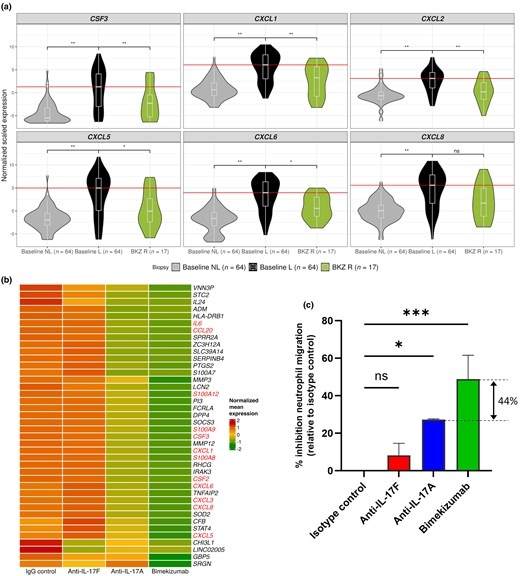
Figure 2. Bimekizumab (BKZ) modulates the upregulation of neutrophil-related genes in hidradenitis
https://doi.org/10.1093/bjd/ljae442
suppurativa (HS) lesional skin. A 2026 study published in Cytokine provides another key piece of evidence. In human dermal fibroblasts, IL-17A is indeed more potent than IL-17F when assessed individually. However, this difference disappears in the presence of TNF. Through synergistic interactions with TNF, IL-17F amplifies inflammatory responses to levels comparable to those induced by IL-17A plus TNF, driving robust upregulation of the neutrophil chemoattractant CXCL8 and tissue-destructive enzymes [3]. These findings suggest that in TNF-rich inflammatory microenvironments in vivo, IL-17F may contribute far more to disease pathology than its standalone activity would suggest.
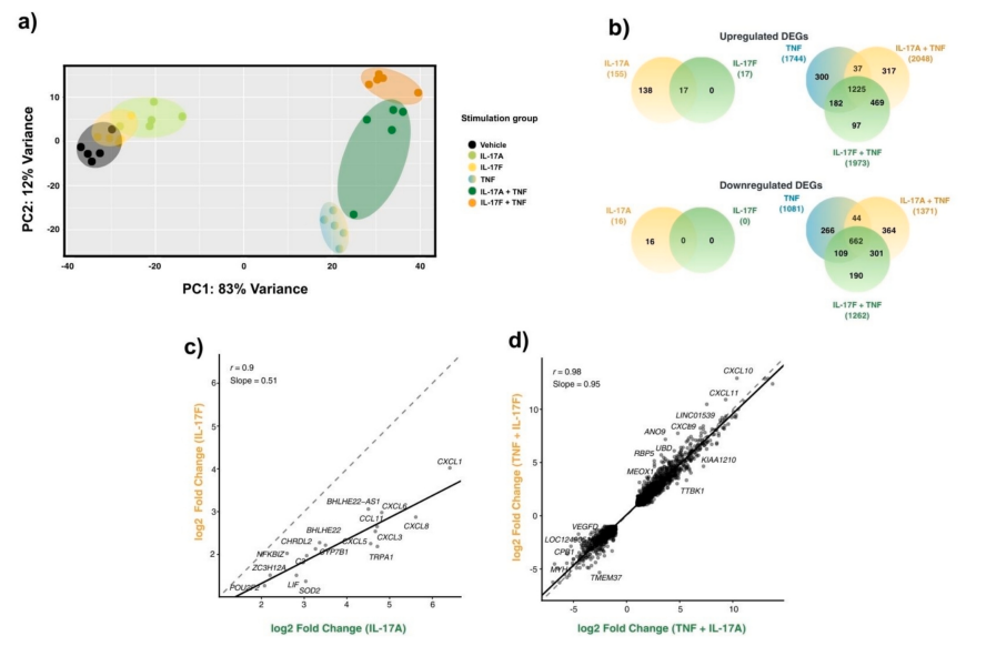
Figure 3. TNF potentiates the transcriptional response of IL-17F to levels comparable to IL-17A in human dermal fibroblasts.
https://doi.org/10.1016/j.cyto.2026.157147
Research in the IL-17 field is shifting from an IL-17A-centric approach toward a more balanced focus on both IL-17A and IL-17F. A 2026 review published in the Mediterranean Journal of Rheumatology highlights that IL-17F is not merely a bystander, but an active contributor to chronic, persistent inflammation and treatment escape. Dual IL-17A/IL-17F inhibition is emerging as an important strategy to overcome the limitations of current therapies and achieve deeper, more durable remission (Figure 4) [4]. Clinical validation of dual-target inhibition is also accelerating worldwide. Bimekizumab has received regulatory approval for psoriasis, psoriatic arthritis, axial spondyloarthritis, and hidradenitis suppurativa. Results from the BE BOLD study announced in March 2026 further established bimekizumab as the first biologic to demonstrate head-to-head superiority over the IL-23 inhibitor risankizumab in psoriatic arthritis, achieving superiority on ACR50 response at Week 16. In the BE HEARD I and II studies, HiSCR50 responses achieved at Week 16 were maintained through Week 48, while three-year follow-up data showed continued improvement in clinical response over time. Sonelokimab, an IL-17A/IL-17F-targeting nanobody, has also demonstrated promising results. In 52-week Phase III data from the VELA program announced in June 2026, 67.2% of patients with hidradenitis suppurativa achieved HiSCR75 and 33.1% achieved HiSCR100, exceeding outcomes reported in previous studies of similar therapies. In the Phase III IZAR-1 trial of sonelokimab in psoriatic arthritis, 42.1% of biologic-naïve patients achieved an ACR50 response at Week 16, and all key secondary endpoints were met.
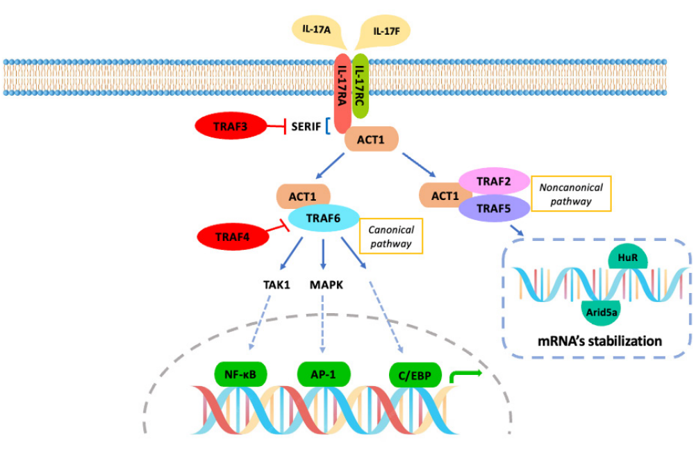
Figure 4. IL-17A/F signalling pathway.
https://doi.org/10.31138/mjr.020925.aar
From Mechanism to Measurement: Evolving Biomarker Requirements for Dual-Target Therapies
The evidence outlined above points to a clear conclusion: IL-17A and IL-17F act in concert to sustain chronic inflammation, and inhibiting both cytokines may offer more complete and durable disease control than targeting IL-17A alone. This shift is beginning to reshape the therapeutic landscape. On August 25, 2026, Laikangqitomab injection (Libeitan®), developed through a collaboration between Livzon MAB and Xinkanghe, received approval from China’s National Medical Products Administration (NMPA). The approval marked a milestone as the first domestically developed biologic in China designed to inhibit both IL-17A and IL-17F. The therapy is indicated for adults with moderate-to-severe plaque psoriasis who are candidates for systemic therapy or phototherapy. In June 2026, its marketing application for ankylosing spondylitis was also accepted for review, further expanding its potential clinical reach. Beyond this approval, multiple dual-target and multitarget candidates are advancing through clinical and preclinical development, reflecting growing momentum across the field (Table 1).
Table 1. Development Pipeline of IL-17A & IL-17F Bispecific and Multispecific Antibodies
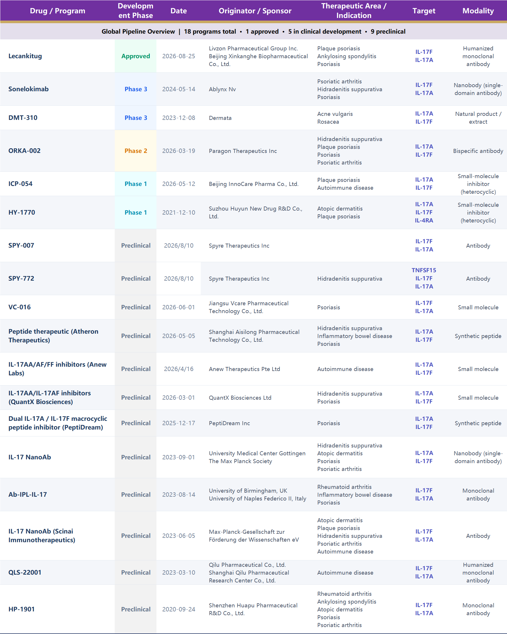
Data as of October 14, 2026
However, dual-target therapies introduce substantially greater complexity in biomarker assessment than single-target approaches. When a drug targets both IL-17A and IL-17F, monitoring IL-17A alone is no longer sufficient to capture the full pharmacodynamic profile of treatment. Accurate characterization requires quantifying all three dimeric forms—IL-17A/A, IL-17F/F, and IL-17A/F—to assess target engagement comprehensively. Distinguishing patient subgroups whose disease is predominantly driven by IL-17A versus IL-17F is also critical to understanding differences in treatment response. More broadly, biomarker assessment must extend beyond individual IL-17 targets to encompass the interconnected Th17 signaling network.
Biomarker Testing: From Single-Target Analysis to Multidimensional Pathway Profiling
In the era of dual-target therapies, biomarker testing is evolving beyond IL-17A-only measurement toward comprehensive pathway-level profiling. As illustrated by the Th17 cell differentiation and plasticity network (Figure 5), inflammatory regulation in autoimmune diseases involves multiple interacting mediators, supporting the development of multiplex biomarker panels.
Upstream regulators: IL-23, IL-1β, IL-6, and TGF-β are key drivers of Th17 cell differentiation and expansion. IL-12/IL-23 p40, a subunit shared by IL-12 and IL-23, provides insight into the activation of both Th1 and Th17 pathways. Core effector cytokines: Biomarker panels should incorporate quantitative measurements of all three IL-17 dimeric forms—IL-17A/A, IL-17F/F, and IL-17A/F—alongside IL-22. IL-17A and IL-17F can form heterodimers in activated CD4⁺ T cells. Quantifying each dimeric form separately is therefore important for characterizing target engagement and assessing the pharmacodynamic effects of dual-target therapies. IL-22, which is also produced by Th17 cells, contributes to epithelial inflammation and the regulation of barrier function. Downstream inflammatory mediators: IL-17 promotes neutrophil recruitment by inducing chemokines such as IL-8/CXCL8. Serum TARC/CCL17 levels are closely associated with disease activity in atopic dermatitis, while IL-31 plays a direct role in itch signaling. Multiplex biomarker testing can provide a more comprehensive picture of inflammatory activity in autoimmune diseases while generating system-level data to support pharmacodynamic assessment, patient stratification, and biomarker monitoring in clinical trials of dual-target therapies [5].
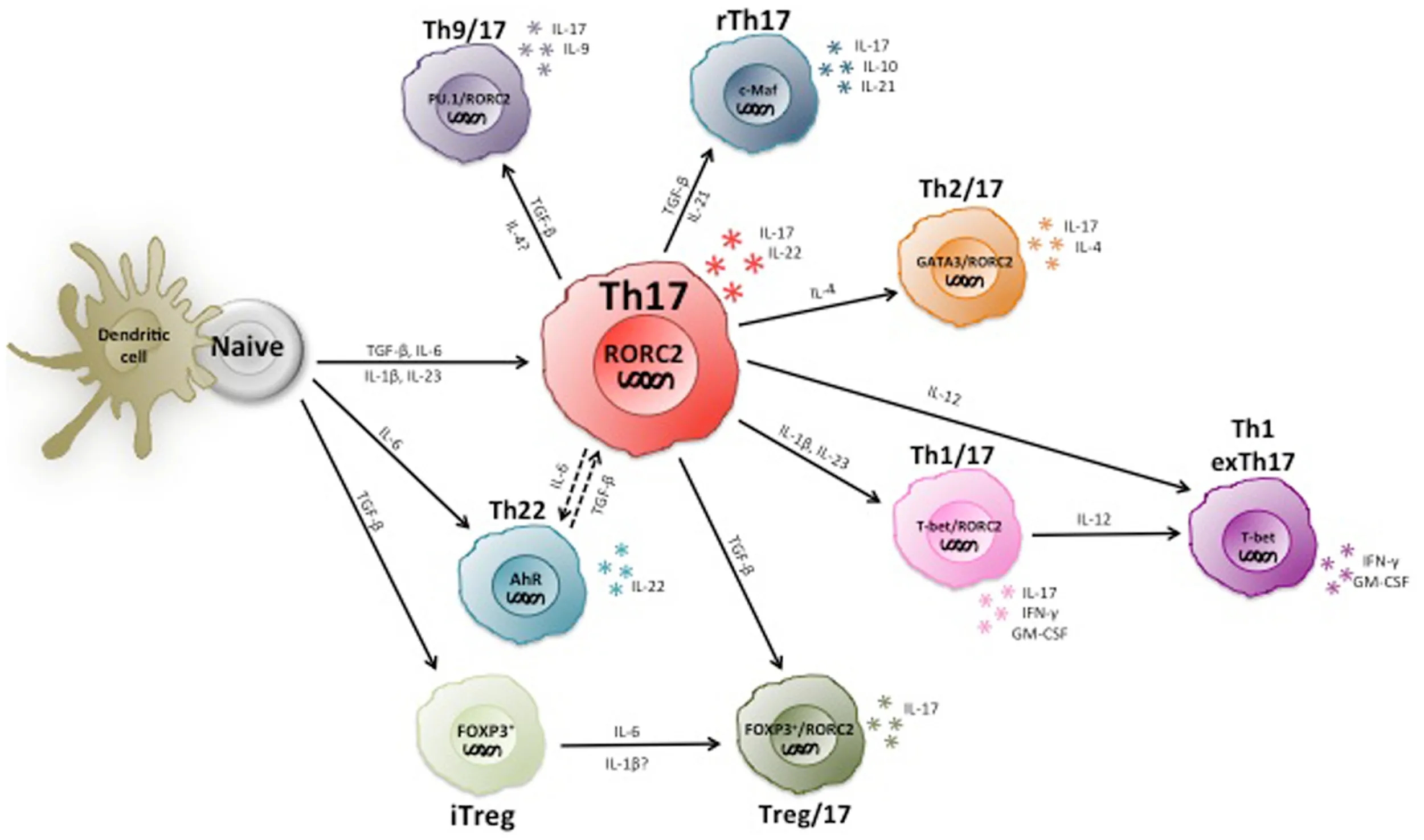
Figure 5. Heterogeneity and plasticity of human Th17 cells.
https://doi.org/10.3389/fimmu.2014.00630
ACROBiosystems Solutions: Comprehensive Biomarker Testing for Autoimmune Diseases
ACROBiosystems and its immunoassay brand, ACRODiagnostic, offer a portfolio of ready-to-use biomarker ELISA kits, rigorously validated in biological samples to support the quantitative analysis of key biomarkers associated with autoimmune diseases, including those discussed above.
| Cat. No. | Product Description | Species |
|---|---|---|
| CEA-C092 | Human IL-17A / CTLA8 ELISA Kit | Human |
| CEA-M272 | Mouse IL-17A ELISA Kit New | Mouse |
| CEA-C233 | Human IL-17F ELISA Kit New | Human |
IL-17A/F ELISA Kits — Coming Soon!
Designed for the quantitative measurement of IL-17A and IL-17F in human serum, plasma, and cell culture supernatants, these assays support antibody drug screening, pharmacodynamic assessment, and biomarker monitoring in clinical trials. To support comprehensive pathway-level profiling in dual-target drug development, we also offer a portfolio of ELISA kits covering key upstream regulators and downstream effectors of the Th17 pathway, including IL-23 (Cat. No. CEA-C093), IL-22 (Cat. No. CEA-C235), IL-12/IL-23 p40 (Cat. No. CEA-C203), IL-6 (Cat. No. CRS-B001), IL-8 (Cat. No. CRS-B004), IL-1β (Cat. No. CRS-B002), TARC/CCL17 (Cat. No. CEA-C029), and IL-31 (Cat. No. CEA-C243). Together, these assays support multiplex biomarker profiling and patient stratification to advance dual-target therapeutic development. Interested in learning more? Contact us to explore the right biomarker testing solutions for your research~ order@acrobiosystems.com
Verification Data
Human IL-17F ELISA Kit(Cat. No. CEA-C233)· Sample Values
Peripheral blood mononuclear cells from a single donor (PBMCs; seeded at 1 x 10⁶/mL) were cultured in RPMI 1640 supplemented with 10% fetal bovine serum, 2 mM L-glutamine, 100 U/mL penicillin, and 100 ug/mL streptomycin sulfate. Cells were left untreated or treated with 10ug/mL of PHA for 1, 3 and 5days.

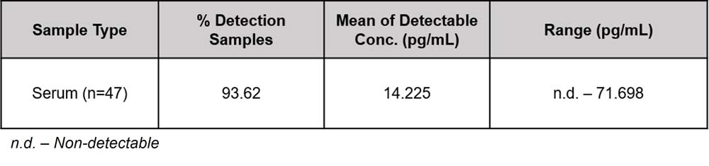
Mouse IL-17A ELISA Kit(Cat. No. CEA-M272)· Sample Values
Forty-seven mouse serum samples were evaluated for the concentrations of mouse IL-17A in assay. The detection IL-17A rate of the samples was 93.62%, the range was ND-71.698 pg/mL, and the mean detectable value was 14.225 pg/mL.
Human IL-17A / CTLA8 ELISA Kit (Cat. No. CEA-C092)·Sample Values
Cell Culture Supernatant 1: Human peripheral blood mononuclear cells (PBMCs) obtained were plated at a density of 1 × 10⁶ cells/mL in complete RPMI 1640 medium containing 10% fetal bovine serum, 2 mM L-glutamine, and antibiotic supplements (penicillin 100 U/mL with streptomycin sulfate 100 μg/mL). Following a 5-day incubation period, experimental groups were either maintained as untreated controls or stimulated with 10 μg/mL phytohemagglutinin (PHA). Subsequent analysis involved quantitative measurement of IL-17A concentrations in collected culture supernatants using standardized immunoassays.