


 Oct 15, 2026
 Page Views:86



From Target Biology to Diagnostic Antibodies: Unpacking the “First Mile” of B7-H3 Development
A pivotal stage in the global development of B7-H3 ADCs is now approaching. On October 10, 2026, Daiichi Sankyo and Merck's I-DXd (DS-7300a, ifinatamab deruxtecan) is set to reach its FDA target action date, potentially marking a new inflection point as the B7-H3 ADC field moves from regulatory review toward commercialization. At the same time, new late-breaking data from multiple Phase 3 B7-H3 ADC studies are slated for presentation at the WCLC 2026 Presidential Symposium, as biopharmaceutical companies worldwide accelerate registration-enabling clinical programs and commercialization strategies.
Once regarded alongside PD-L1 as a next-generation immuno-oncology target, B7-H3 is rapidly advancing toward the commercial stage. For researchers and biopharmaceutical companies developing B7-H3-targeted therapies, one critical upstream consideration has remained integral throughout the development-to-market pathway: B7-H3 IHC testing. Why has protein-level assessment become such an important step in B7-H3 drug development? This article examines the path from target biology to diagnostic antibodies, unpacking the “first mile” of B7-H3 drug development and exploring the role of IHC in translating B7-H3-targeted therapies from discovery and clinical development toward commercialization.
What Is B7-H3? A Dual-Function Target with Both Immunosuppressive and Non-Immune Oncogenic Roles
1. Structural Features: Two Isoforms and a Soluble Form
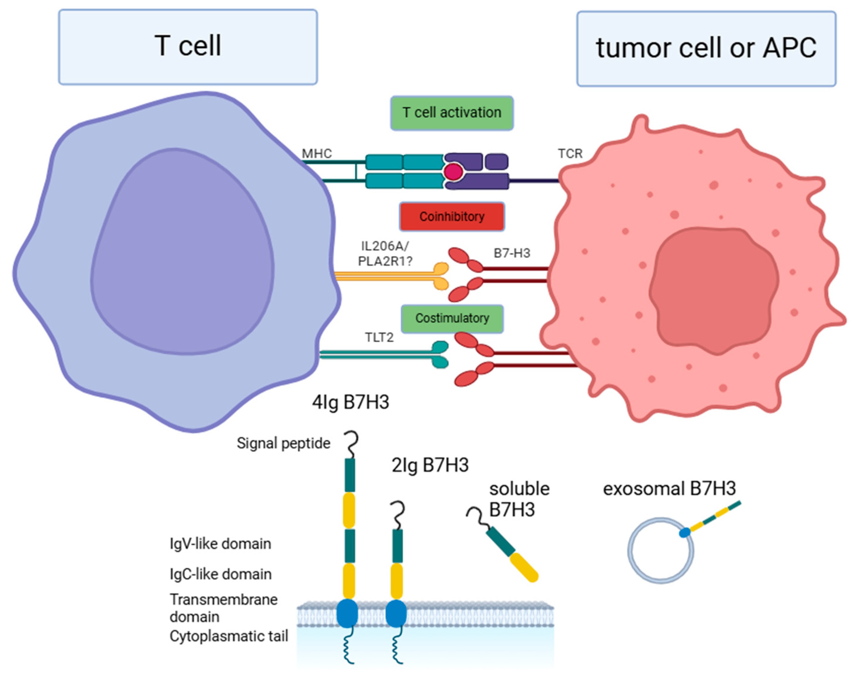
B7-H3 (CD276) is a type I transmembrane protein belonging to the B7 family of immune checkpoint molecules. It was first identified in 2001. In humans, B7-H3 exists in two isoforms: 2IgB7-H3, containing one pair of IgV-like and IgC-like domains, and 4IgB7-H3, containing two identical pairs of these domains. The 4IgB7-H3 isoform is predominantly expressed on the surface of human monocytes and tumor cells. A soluble form of B7-H3 (sB7-H3) is also detectable in serum and extracellular vesicles and may arise through proteolytic cleavage or alternative splicing of 2IgB7-H3[1]. This structural complexity underscores an important limitation of mRNA-based analysis alone: transcript abundance may not accurately reflect B7-H3 protein expression at the cellular level.
https://doi.org/10.3390/cells14070530
Figure 1. B7H3 structure and its putative receptor.
2. Expression Profile: High Tumor Expression with Limited Expression in Normal Tissues
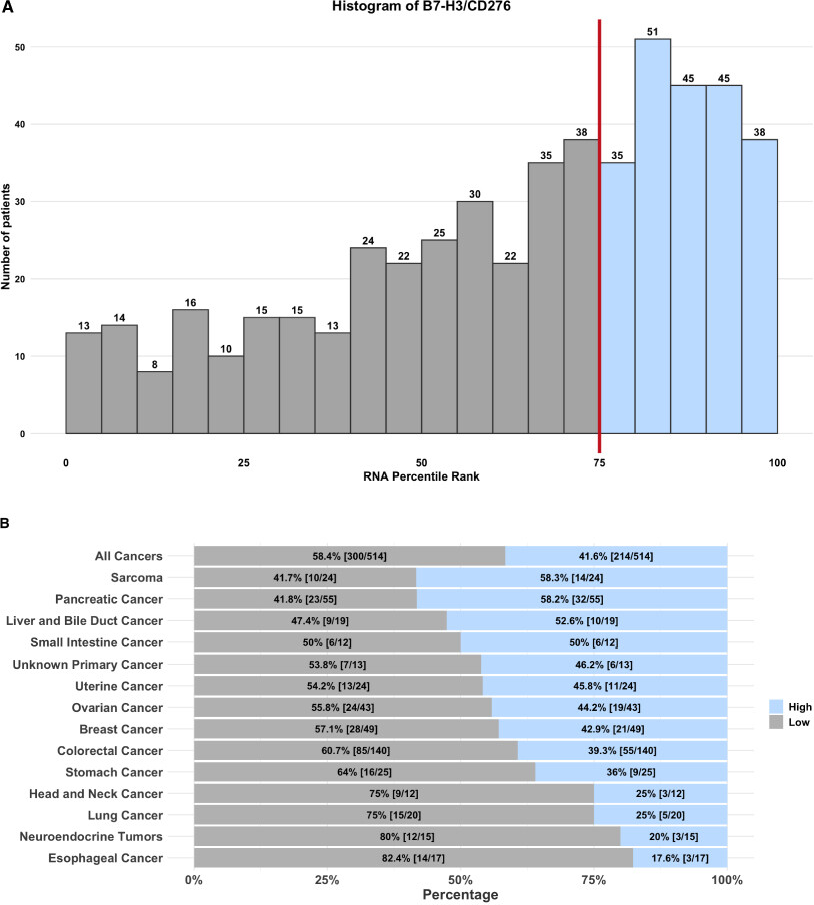
One of the most compelling features of B7-H3 expression is its abundant expression in malignant cells coupled with minimal expression in healthy tissues. This “high in tumors, low in normal tissues” expression profile provides a key biological rationale for B7-H3 as an attractive ADC target. Larkin et al. conducted a comprehensive analysis of B7-H3 RNA expression in tumor samples from 514 patients with advanced cancers treated at the Moores Cancer Center at the University of California San Diego. The analysis showed that 41.6% of patients exhibited high levels of B7-H3 expression. B7-H3 positivity is also substantial across gastrointestinal malignancies, with reported positive expression rates ranging from 32%–87% in colorectal cancer, 39.4%–69.2% in gastric cancer, 30.7%–97.5% in hepatocellular carcinoma, 41.2%–77.8% in pancreatic cancer, and 55.8%–69.7% in esophageal cancer[2].
https://doi.org/10.2147/ITT.S534666
Figure 2 Proportion of patients exhibiting high B7-H3/CD276 expression.
3. Dual Mechanisms: Immune Evasion and Tumor-Promoting Activity
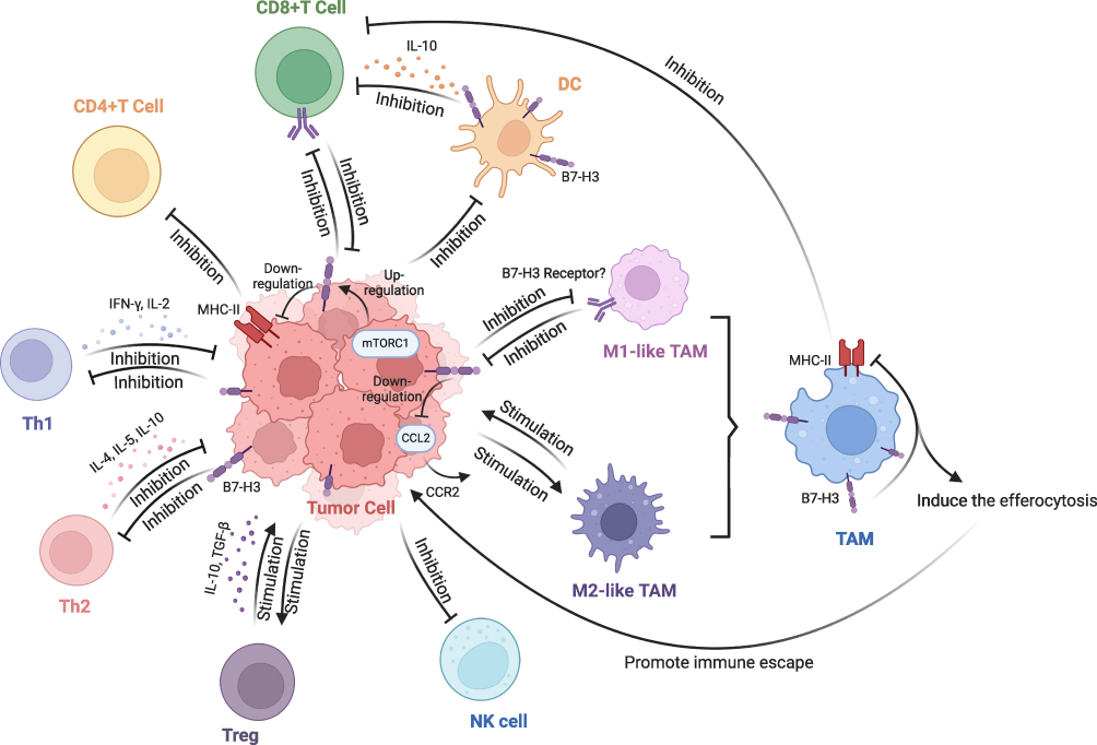
A defining feature of B7-H3 is its ability to drive both immunosuppressive and non-immune tumor-promoting pathways. Within the tumor microenvironment (TME), B7-H3 contributes to tumor progression through multiple immunomodulatory mechanisms, including dampening T-cell responses, promoting the polarization of tumor-associated macrophages (TAMs) toward an M2-like phenotype, and suppressing the function of other immune cell populations[3]. Beyond its immunoregulatory effects, B7-H3 also exerts non-immune functions that promote tumor cell proliferation, migration, invasion, metabolic dysregulation, angiogenesis, and resistance to therapy. High B7-H3 expression has been associated with poor prognosis, advanced disease stage, and lymph node metastasis. Together, these features have positioned B7-H3 as a key target of interest across multiple therapeutic modalities, including antibody-drug conjugates (ADCs), bispecific antibodies, and CAR-T cell therapies.
https://doi.org/10.2147/ITT.S507522
Figure 3 Immunomodulatory function B7-H3 in the TME.
Why Is IHC Testing a Critical Upstream Gateway in B7-H3 Drug Development?
1. mRNA ≠ Protein: Expression Discordance Driven by Post-Transcriptional Regulation
B7-H3 exists in two splice variants, 2IgB7-H3 and 4IgB7-H3, and substantial discordance has been observed between B7-H3 mRNA and protein expression levels, suggesting active post-transcriptional regulation. mRNA-based assays, including qPCR and RNA-seq, cannot reliably capture the actual level of B7-H3 protein expression or its localization on the tumor cell membrane. This distinction is particularly important for ADCs, whose activity depends on sufficient target-antigen density at the cell surface. These considerations further underscore the critical role of IHC-based protein-level assessment.
2. Limitations of Currently Available Antibodies
In the current B7-H3 IHC landscape, different antibodies have distinct application profiles:
Some commonly used clones are better suited to flow cytometry and frozen tissue sections, with more limited utility in formalin-fixed, paraffin-embedded (FFPE) specimens.
Other antibodies are validated for FFPE applications but typically require working concentrations of approximately 20 μg/mL (around a 1:50 dilution). In large, multicenter clinical trials, this can place greater demands on reagent consumption management and lot-to-lot consistency.
For ADC clinical trials requiring standardized staining across hundreds or even thousands of specimens, these limitations may become practical bottlenecks in assay scalability, reagent management, and analytical consistency.
HGS-S609: A Systematically Validated B7-H3 IHC Antibody
Accurate and reliable IHC antibodies are essential tools for characterizing B7-H3 expression, supporting indication assessment, and evaluating therapeutic response. Selecting a high-performance B7-H3 IHC antibody provides a critical foundation for efficient drug development.
To support the development of B7-H3-targeted therapies, we have launched a new B7-H3 IHC antibody, HGS-S609 (Clone 7A1). This recombinant rabbit monoclonal antibody is compatible with the Leica BOND III automated staining platform and has undergone systematic validation on the ACRODiagnostic pathology platform.

In addition to B7-H3, we are expanding our portfolio to include other key targets within the B7 family. Our recombinant rabbit monoclonal antibody against B7-H1 (PD-L1), HCS-S240, is now available, while a recombinant rabbit monoclonal antibody against B7-H4 is currently in development. Together, these offerings provide streamlined access to a comprehensive, one-stop solution for B7 family IHC testing.
For gastrointestinal cancers, including colorectal, pancreatic, and gastric cancers, we also offer IHC antibodies targeting a broad range of clinically relevant diagnostic biomarkers, these products provide a comprehensive suite of diagnostic reagent tools to support B7-H3-targeted drug development as programs advance into the clinical stage.
Explore More IHC Products
Validation Data
1. Comparative Multi-Tissue IHC Staining: HGS-S609 vs. Company A
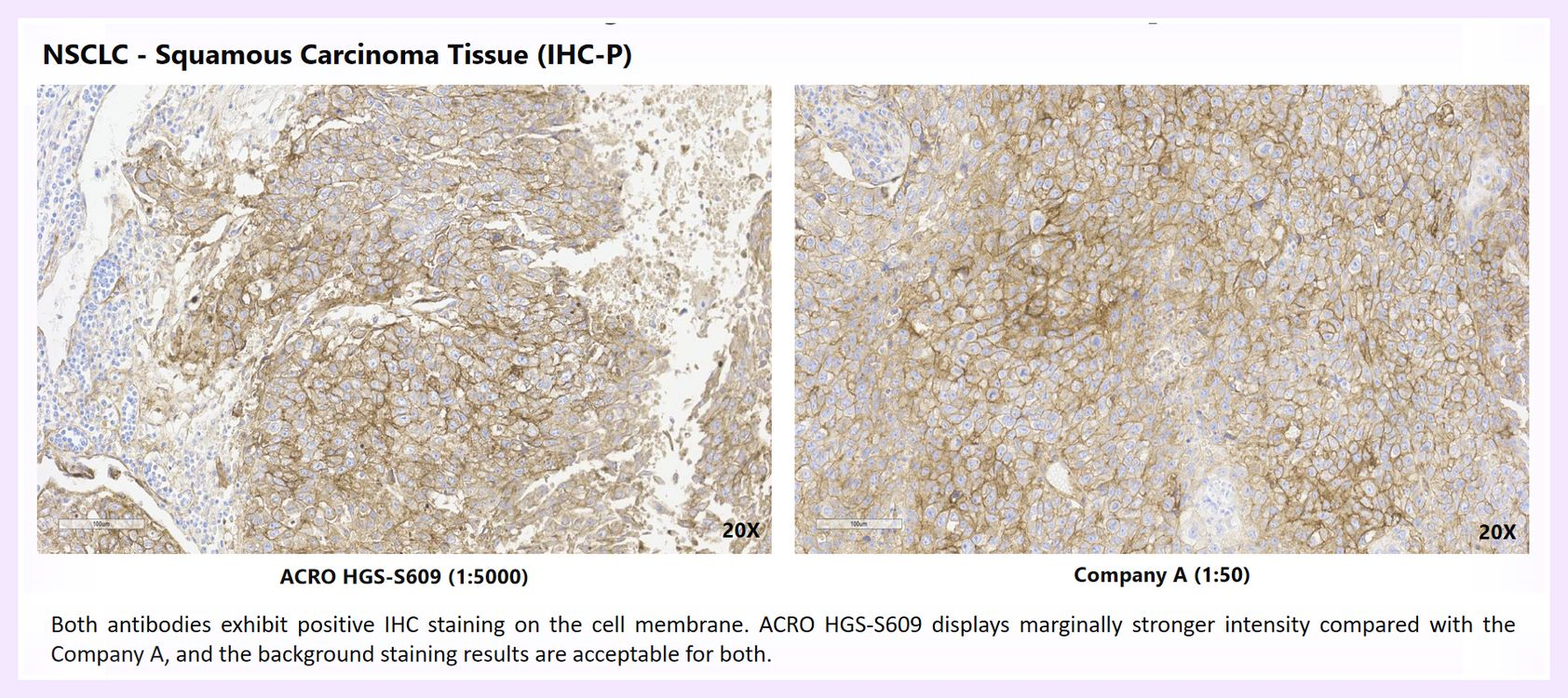
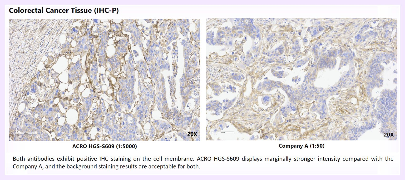
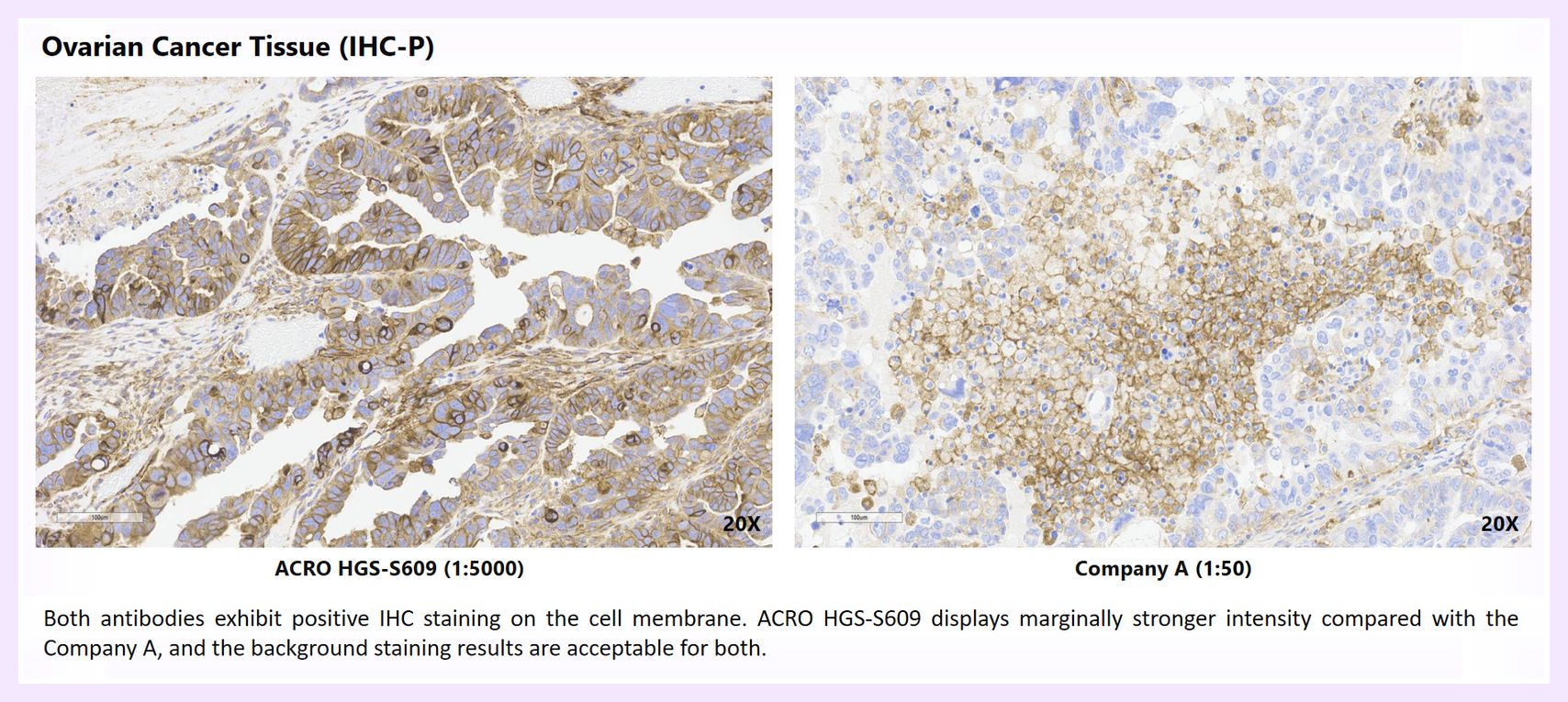
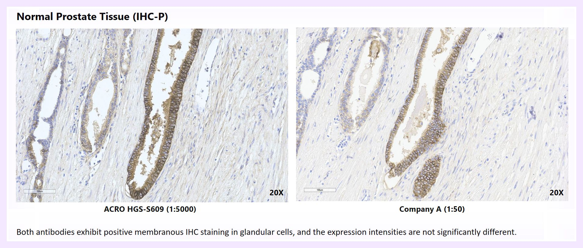
HGS-S609 has been analytically validated across a broad range of tissue types, including two normal control tissues (prostate and placenta) and multiple tumor types, such as squamous cell carcinoma of the lung, lung adenocarcinoma, colorectal cancer, prostate cancer, and ovarian cancer. This broad tissue coverage provides a strong foundation for patient selection and biomarker analysis across different indications in B7-H3 ADC clinical development. Selected staining results are shown below.
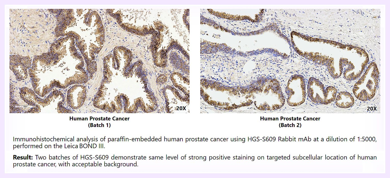
2. Batch-to-Batch Consistency Validation
Immunohistochemical analysis was performed on paraffin-embedded human prostate cancer tissue using two batchs of HGS-S609 at a 1:5,000 dilution on the Leica BOND III automated staining platform. The results demonstrated comparable positive staining intensity and consistent subcellular localization across both antibody lots in human prostate cancer tissue, with acceptable background staining observed for both lots.