


 Sep 18, 2026
 Page Views:138


Introduction
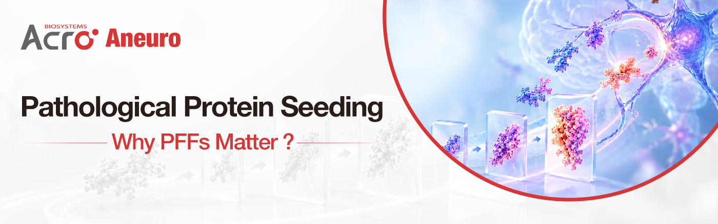
In neurodegenerative disease research, misfolded proteins can act as pathological seeds, recruiting native proteins into aggregates that propagate across cells and brain regions. This seeding process is a key feature of proteinopathies involving α-Synuclein (α-Syn), Tau, and APP, and is widely studied in Parkinson's disease (PD), Alzheimer's disease (AD), and related neurodegenerative disorders.
To model these processes experimentally, researchers often use pre-formed fibrils (PFFs) as defined pathological seeds to initiate protein aggregation and propagation in cellular, organoid, or animal models.
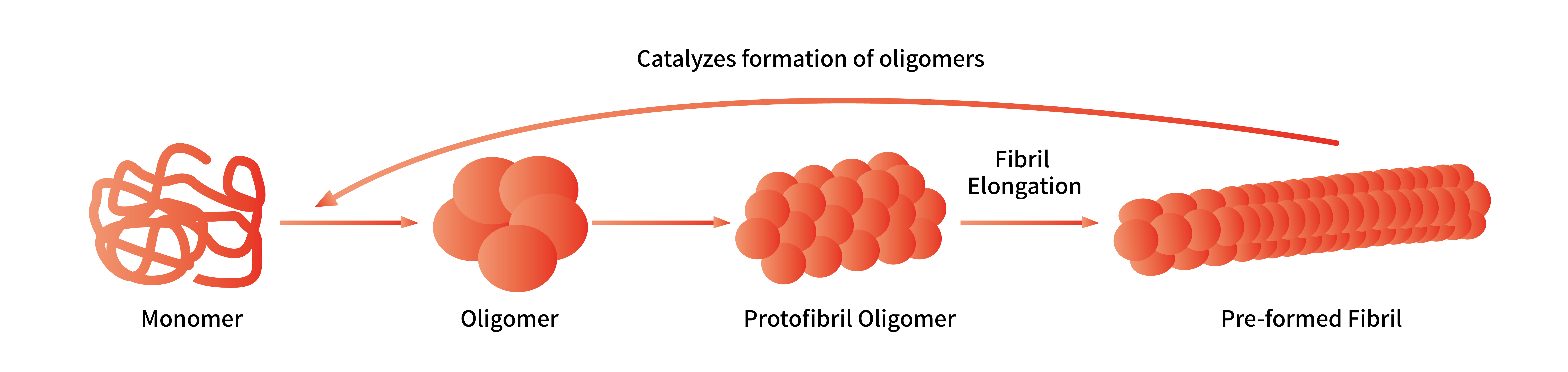
Schematic of the PFFs Formation Process
PFFs in Neurodegenerative Disease Research: From Mechanistic Studies to Therapeutic Evaluation
In recent years, from mechanistic dissection to disease modeling and therapeutic validation, PFFs are increasingly being used in high-impact neuroscience studies, from mechanistic investigations and disease modeling to therapeutic evaluation.
Identifying a Key Mediator of α-Syn Fibril Uptake
A 2025 Science study addressed a central question: how do neurons take up pathological α-Syn?
The study found that the PD risk gene FAM171A2 promotes the endocytosis of α-Syn fibrils and further influences the spread of α-Syn pathology and neurotoxicity. Researchers inoculated recombinant α-Syn PFFs into the mouse striatum and observed that PFFs were taken up by axon terminals, accompanied by the gradual accumulation of α-Syn pathology in distant brain regions. Further experiments showed that increasing FAM171A2 expression exacerbated PFF-induced pathology, while reducing its expression was protective. Notably, the recognition of PFFs by FAM171A2 was not a simple "bind-any-α-Syn" mechanism: its extracellular domain had a significantly higher affinity for α-Syn fibrils than for monomeric α-Syn.
This study demonstrates that PFFs are a vital tool for dissecting how pathological proteins enter neurons, how they propagate, and which molecules participate in the process.
Specific Fibril Conformations Can Be Faithfully Propagated In Vivo
A 2025 Nature study focused on a specific fibril conformation (1B) generated in vitro.
When injected into mouse brains, these fibrils self-replicated in vivo, inducing glial cytoplasmic inclusions (GCIs) characteristic of multiple system atrophy (MSA), which spread over time to multiple brain regions. Critical evidence came from structural validation: the atomic structure of newly formed fibrils (1BP) purified from the brains of affected mice was highly consistent with that of the originally inoculated 1B fibrils. Passage experiments further confirmed that brain homogenates containing 1BP, when inoculated into new mice, could still replicate the same MSA-like pathology.
This indicates that the specific conformation of fibrils determines the type and fidelity of the induced pathology. For the same protein, different PFF conformations can give rise to distinct pathological phenotypes, and this conformational information remains highly stable during replication.
PFF-based Models Enable Evaluation of Tau Clearance Strategies
A 2026 Nature Biomedical Engineering study applied PFFs in the therapeutic validation stage.
In a cellular model, Tau PFFs first induced reproducible insoluble phosphorylated Tau pathology, providing a standardized pathological starting point for evaluating clearance strategies. Subsequently, using the pathological model established by Tau PFFs, the researchers confirmed that engineered autophagy receptors fused with LC3A and delivered via small extracellular vesicles (sEVs) significantly reduced PFF-induced phosphorylated Tau. Furthermore, in TauP301S mice, this strategy also effectively reduced insoluble phosphorylated Tau in brain tissue.
By providing a defined and reproducible pathological starting point, PFF-based models enable systematic evaluation of therapeutic strategies designed to reduce or reverse Tau pathology.
Cellular Environments Can Enhance the Seeding Activity of Synthetic Aβ
A 2016 Journal of Neuroscience study asked: how can synthetic Aβ acquire seeding activity more similar to that of brain-derived seeds?
Researchers established a long-term hippocampal slice culture model, continuously adding synthetic Aβ to the culture medium along with a single addition of brain extract from APP transgenic mice. After several weeks, abundant Aβ deposits formed in the slices, accompanied by microglial activation, dendritic spine loss, and other pathologies. Fixed, inactivated slices showed only minimal marginal deposits, suggesting that the living cellular environment is a crucial condition for this conversion process. Further re-implantation experiments showed that homogenates from slices containing Aβ deposits, when injected into the brains of APP transgenic mice, induced pronounced amyloid-β plaque formation, with seeding activity significantly higher than that of synthetic Aβ aggregated in vitro alone.
This demonstrates that the living cellular environment promotes seed-induced conversion of synthetic Aβ, endowing it with stronger in vivo seeding activity.
Aneuro: Well-Characterized PFFs for Reproducible Disease Modeling
Aneuro, the neuroscience-focused brand of ACROBiosystems, has developed a PFF product portfolio targeting key proteins including Tau, α-Synuclein, and APP.
Product List
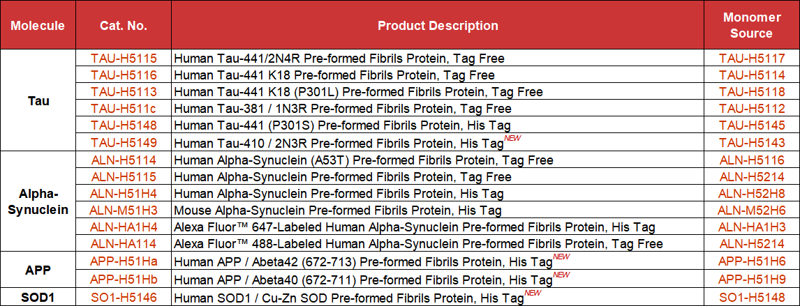
Effective PFFs must do more than form fibrillar structures—they need to function as consistent pathological seeds capable of inducing disease-relevant phenotypes. To this end, we have performed multi-level validation—from assembly morphology and recruitment activity to cellular induction efficiency and organoid pathology modeling—to confirm the structural and functional performance of our PFFs.
Key Validation Data
PFF Morphology Characterized by Electron Microscopy
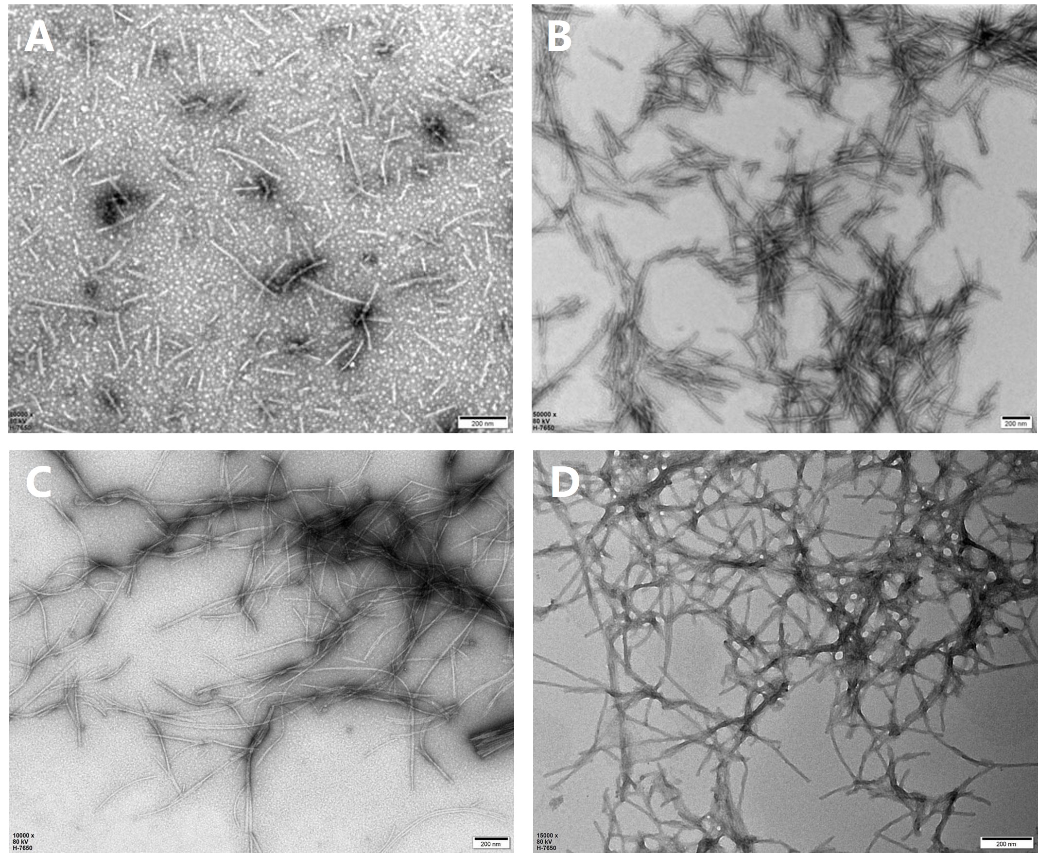
Transmission electron microscopy (TEM) was used to observe the morphological characteristics of wild-type and mutant Tau PFFs and α-Syn PFFs. Negative-stain TEM showed that both wild-type and mutant Tau PFFs and α-Syn PFFs presented as fibrillar structures with diameters between 100 and 500 nm. Most fibrils were straight, with occasional twisting. (A) Tau-441/2N4R PFFs (Cat. No. TAU-H5115); (B) Tau-441 K18 (P301L) PFFs (Cat. No. TAU-H5113); (C) α-Syn PFFs (Cat. No. ALN-H5115); (D) α-Syn (A53T) PFFs (Cat. No. ALN-H5114); scale bar: 200 nm.
ThT Fluorescence Assay Confirms PFFs Seeding Activity
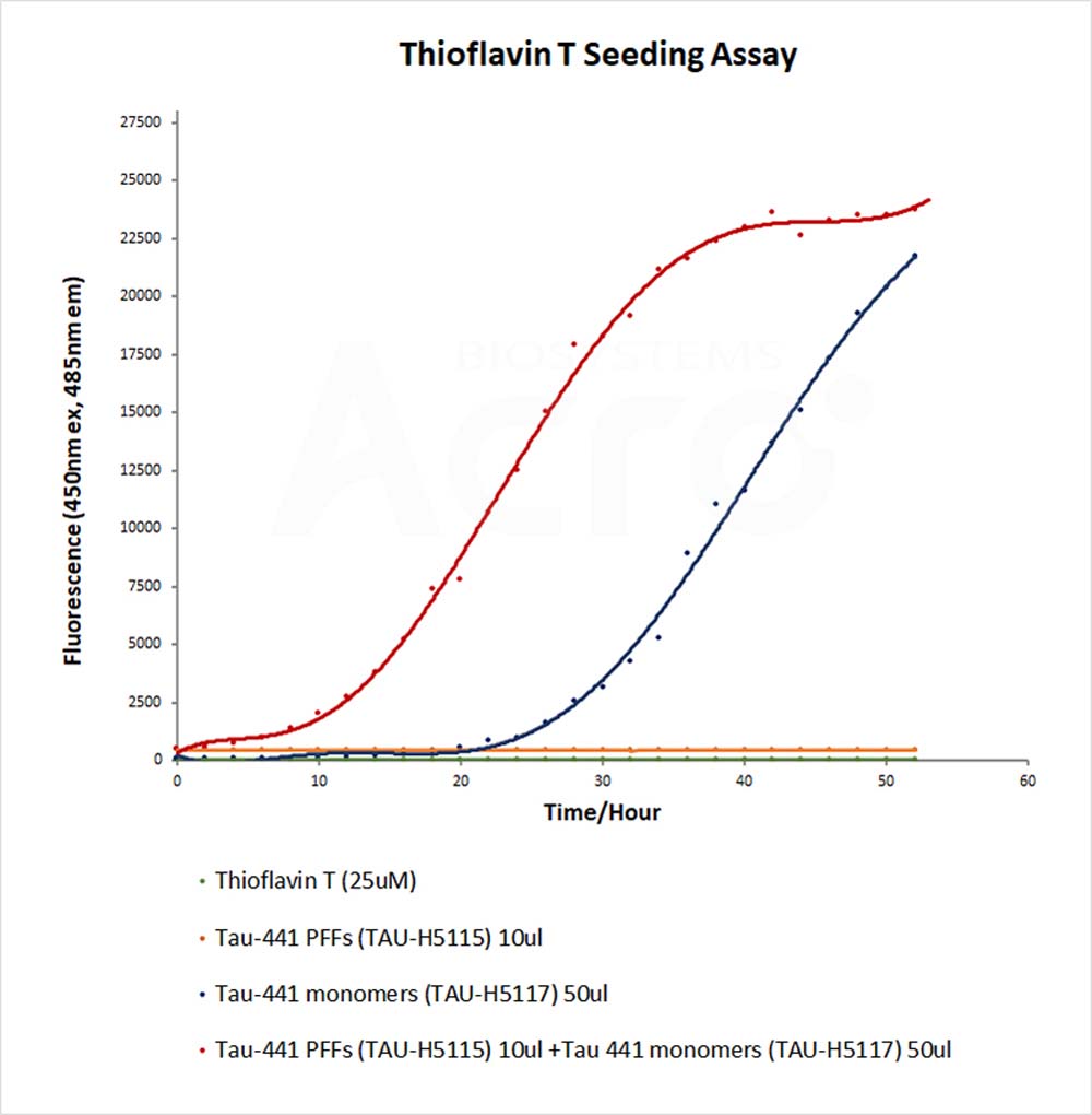
Thioflavin T (ThT) is a fluorescent dye that binds to β-sheet-rich structures and enhances fluorescence. In the Tau ThT assay, Tau monomer (blue curve) aggregated slowly, with fluorescence beginning to rise after 25 hours. The addition of Tau PFFs (red curve) significantly accelerated aggregation, shortening the lag phase and increasing fluorescence intensity. No significant fluorescence change was observed for Tau PFFs alone (orange curve).
Cell-Based Assay Confirming High Inductive Activity of PFFs
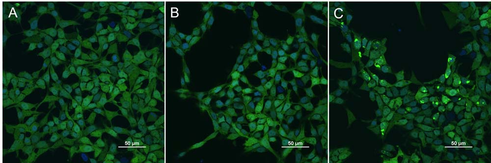
Confocal microscopy revealed obvious Tau protein aggregation in a Tau-K18 (GFP) stable cell line (Cat. No. CHEK-ATP087) induced by Tau PFFs. (A) Transfection with Lipo2000; (B) Co-transfection with Lipo2000 and Tau monomer protein; (C) Co-transfection with Lipo2000 and Tau PFFs.
PFFs for Organoid Neurodegenerative Disease Modeling
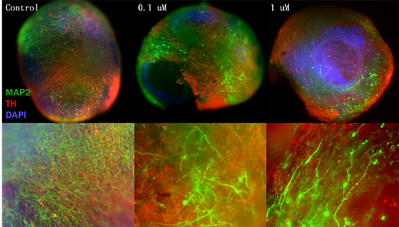
In a PD model, the addition of α-Syn PFFs disrupted the expression of MAP2 and TH, indicating that mature neurons (MAP2) and dopaminergic neurons (TH) were damaged by α-Syn PFFs.
Aneuro: Advance Neuroscience Research
Beyond PFFs, Aneuro offers target proteins, cell lines, p-Tau antibodies, and other research tools designed to support neuroscience research and therapeutic development.