



 Sep 3, 2026
 Page Views:247


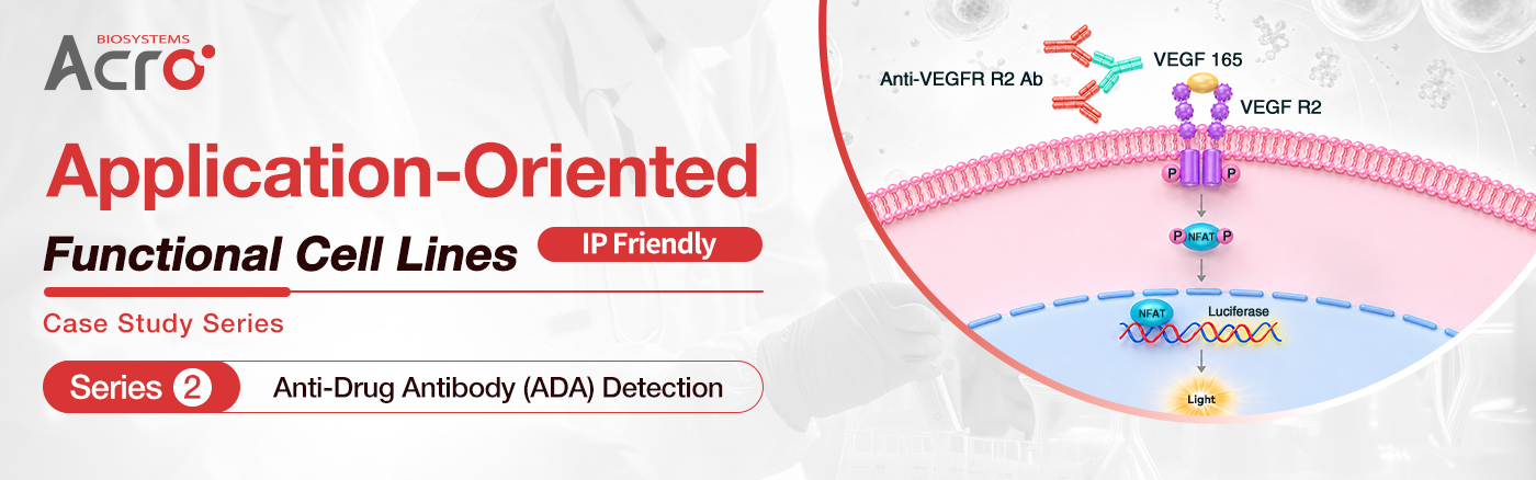
Application‑Oriented Functional Cell Lines | Case Study Series
During drug development, functional cell lines are critical tools connecting drug design, in vitro functional validation, and clinical translational prediction, forming an essential foundation for drug efficacy assessment.
For drug development researchers, ideal cell lines feature qualified performance metrics and are engineered for practical R&D needs. They precisely recapitulate drug's mechanism of action (MOA), and fit multiple assay scenarios, and generate stable, reliable data as well as regulatory-supporting evidence to guide research decisions.
Focused on real-world drug discovery workflows, ACROBiosystems is committed to delivering "Application-Oriented Functional Cell Lines". Built around practical application demands, our stable, reliable, and reproducible cell line models help deliver trustworthy experimental results and accelerate drug development.
We will launch a series of application case studies to illustrate how functional cell lines benefit drug development. Content includes assay strategies, product selection, protocols and data analysis, actionable reference solutions for researchers.
📚Previously in this series:
Series 1: Functional Cell Lines for ADCC/ADCP Functional Validation
Series 2: we focus on a critical component of immunogenicity assessment in biopharmaceutical development: anti-drug antibody (ADA) testing.
ADA Detection: A Non-negotiable for Biopharmaceutical Development
Monoclonal antibodies, fusion proteins and other therapeutic proteins may trigger an immune response and induce anti-drug antibody (ADA). ADA can neutralize drug activity and reduce systemic drug exposure, leading to diminished efficacy or even treatment failure. They may also cause safety events such as infusion reactions and hypersensitivity. During clinical immunogenicity assessment and long-term treatment monitoring of therapeutic proteins, it is essential to rapidly determine whether ADA — particularly neutralizing anti-drug antibodies (nADA) — impair drug activity. These data support early warning of loss of response (LOR), optimization of dosing strategies, and mitigation of immunogenicity risk.
The FDA's 2014 guidance, Immunogenicity Assessment for Therapeutic Protein Products, recommends a risk-based strategy for therapeutic protein products—with systematic assessment and control of immunogenicity risk throughout both nonclinical and clinical stages. ADA testing is a key component of immunogenicity risk control. The FDA guidance also incorporates neutralizing activity assays into a comprehensive ADA analysis strategy and emphasizes that assay design should be aligned with the drug's MOA.
Reporter Cell Lines: Bridging Binding Detection and Neutralizing Activity Assessment
Current ADA testing strategies rely primarily on immunoassay-based methods, including enzyme-linked immunosorbent assay (ELISA), electrochemiluminescence (ECL), surface plasmon resonance (SPR), and radioimmunoassay (RIA). These methods can determine whether an ADA binds to the therapeutic proteins, but they cannot determine whether the ADA actually inhibits drug function.
To evaluate the actual impact of nADA on drug activity, the assay must reflect the drug's real MOA; by leveraging reporter cell lines that match the drug's MOA, the downstream biological effect of drug–target interaction is converted into a readable detection signal—completing the transition from "antigen–antibody binding detection" to "neutralization activity assessment."
ACROBiosystems has developed a portfolio of luciferase-based reporter cell lines with clearly defined MOA. These quantitative, cell-based tools address a key limitation of conventional immunoassays: they measure not only binding, but also whether an ADA inhibits drug function.
Using a reporter cell line, a sensitive and reliable nADA assay can be established in three steps:
1. Antigen stimulation: Determine the optimal antigen concentration.
2. Drug neutralization: Determine the optimal therapeutic antibody concentration.
3. nADA detection: Evaluate nADA activity in the test sample.
Application Case Study: Three-Steps nADA Assay Workflow for Bevacizumab nADA Detection
This case study demonstrates a three-step workflow for assessing neutralizing anti-drug antibodies against bevacizumab using a VEGF R2/NFAT luciferase reporter system.
Products Used in This Case Study
Reporter cell line: Human VEGF R2 (Luc) HEK293 Reporter Cell (Cat. No. CHEK-ATF044)
Recombinant protein: Human VEGF165 Protein (Cat. No. VE5-H4210)
Anti-drug Antibody: Anti-Bevacizumab Antibody (Cat. No. BEB-Y12)
Step 1: Determine the Optimal Antigen Concentration
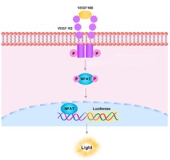
VEGF165 protein binds to the VEGF R2 on the cell surface, activating the downstream NFAT signaling pathway and driving luciferase reporter gene expression to produce a luminescent signal. Higher antigen concentration yields a stronger signal.
Experimental Results: Human VEGF165 protein was serially diluted and co-incubated with the Human VEGF R2 (Luc) HEK293 reporter cell line; relative luminescence units (RLU) were measured across the concentration range. The working antigen concentration for the experiment was determined as EC50 = 10.47 ng/mL.
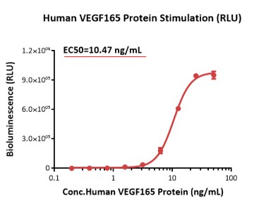
Figure 1. Human VEGF165 Protein Stimulation.
Step 2: Determine the Optimal Therapeutic Antibody Concentration
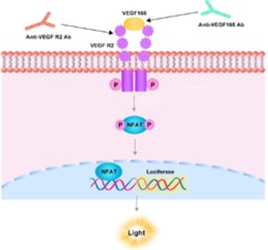
Bevacizumab competitively binds VEGF165 and blocks its interaction with VEGF R2, which inhibits NFAT pathway activation and reduces the luminescent signal.
Experimental Results: Using the VEGF165 concentration established in Step 1 (10.47 ng/mL), bevacizumab was serially diluted and co-incubated with the Human VEGF R2 (Luc) HEK293 reporter cell line; based on the measured RLU values, the working bevacizumab concentration for subsequent nADA detection was determined as EC50 = 0.06299 µg/mL.
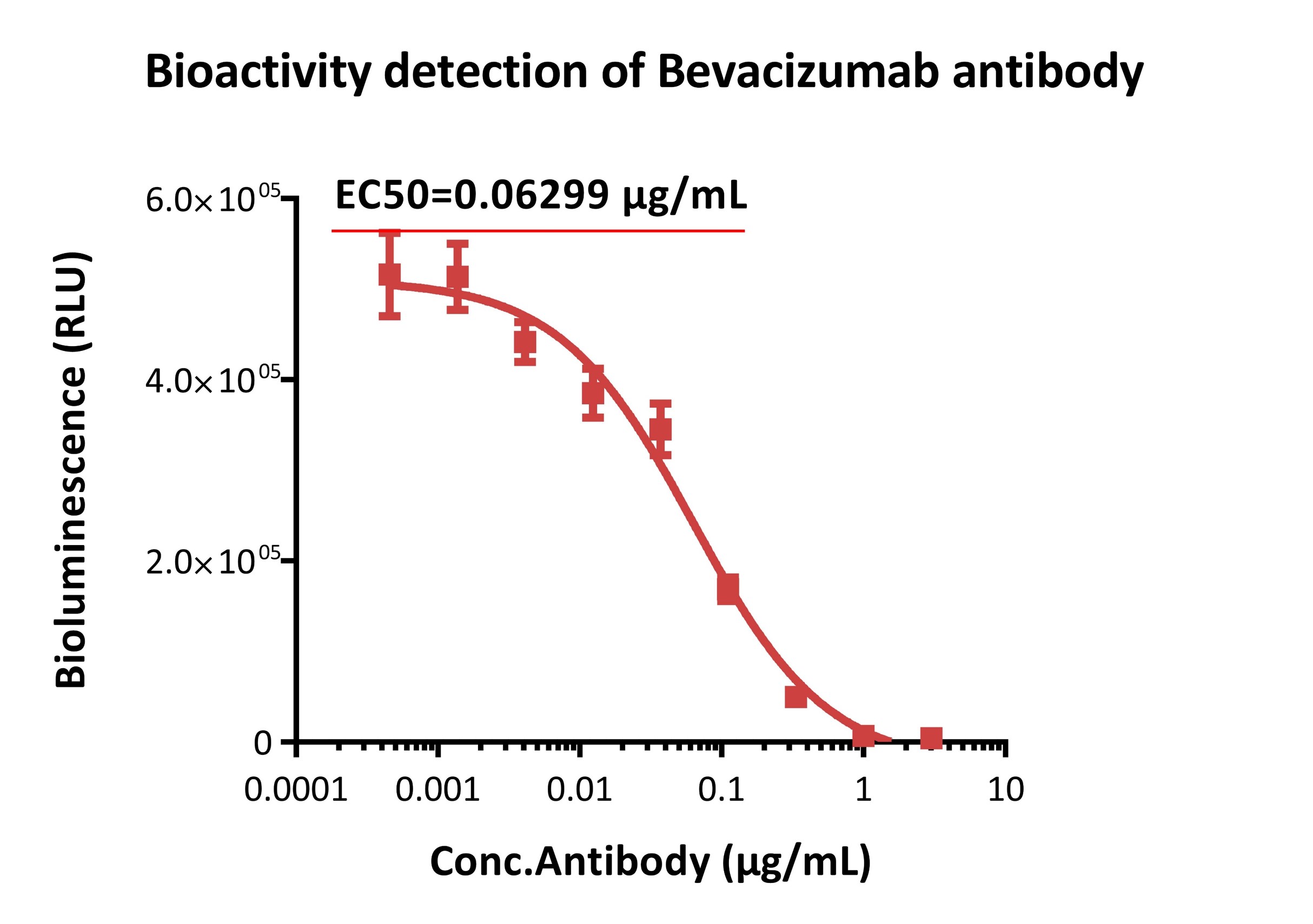
Figure 2. Bioactivity Detection of Bevacizumab.
Step 3: Functional nADA Detection
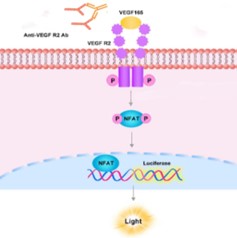
When Bevacizumab nADA is present in a sample, the nADA binds bevacizumab and blocks its neutralization of VEGF165, allowing VEGF165 to re-activate the VEGF R2 signaling pathway and raising the luminescent signal again—the so-called "de-repression" phenomenon—which characterizes the nADA's activity.
Experimental Results: Anti-bevacizumab antibodies were serially diluted to simulate nADA-positive samples and tested at the bevacizumab concentration of 0.06299 µg/mL established in Step 2; the simulated nADA yielded EC50 = 0.5777 µg/mL with a maximum neutralization rate >100%, enabling quantitative characterization of the nADA's functional activity.
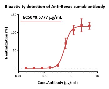
Figure 3. Bioactivity Detection of Anti-Bevacizumab Antibody.
Case Study Summary
This case study provides a complete, three-step nADA detection workflow and demonstrates that ACROBiosystems reporter cell lines can directly characterize the effect of nADA on drug neutralizing activity. The assay delivers sensitive, quantitative readouts, straightforward operation and a reusable experimental system.
Moving forward, we will continue to expand our Application-Oriented Functional Cell Lines Case Study Series covering more drug development scenarios, standardized assay protocols, practical product selection guidance, and real experimental data.
We welcome your feedback on future research topics and technical challenges. Stay tuned for more application-driven case studies tailored to your drug development needs. Explore ACROBiosystems ADA-related products and services.
Explore ACROBiosystems ADA-related Products and Services